


 Foundational characteristics of cancer include proliferation, angiogenesis, migration, evasion of apoptosis, and cellular immortality. Find key markers for these cellular processes and antibodies to detect them.
 The SUMOplot™ Analysis Program predicts and scores sumoylation sites in your protein. SUMOylation is a post-translational modification involved in various cellular processes, such as nuclear-cytosolic transport, transcriptional regulation, apoptosis, protein stability, response to stress, and progression through the cell cycle.
 The Autophagy Receptor Motif Plotter predicts and scores autophagy receptor binding sites in your protein. Identifying proteins connected to this pathway is critical to understanding the role of autophagy in physiological as well as pathological processes such as development, differentiation, neurodegenerative diseases, stress, infection, and cancer.
ATG16L Antibody
Purified Rabbit Polyclonal Antibody (Pab)
- SPECIFICATION
- CITATIONS: 2
- PROTOCOLS
- BACKGROUND

Application
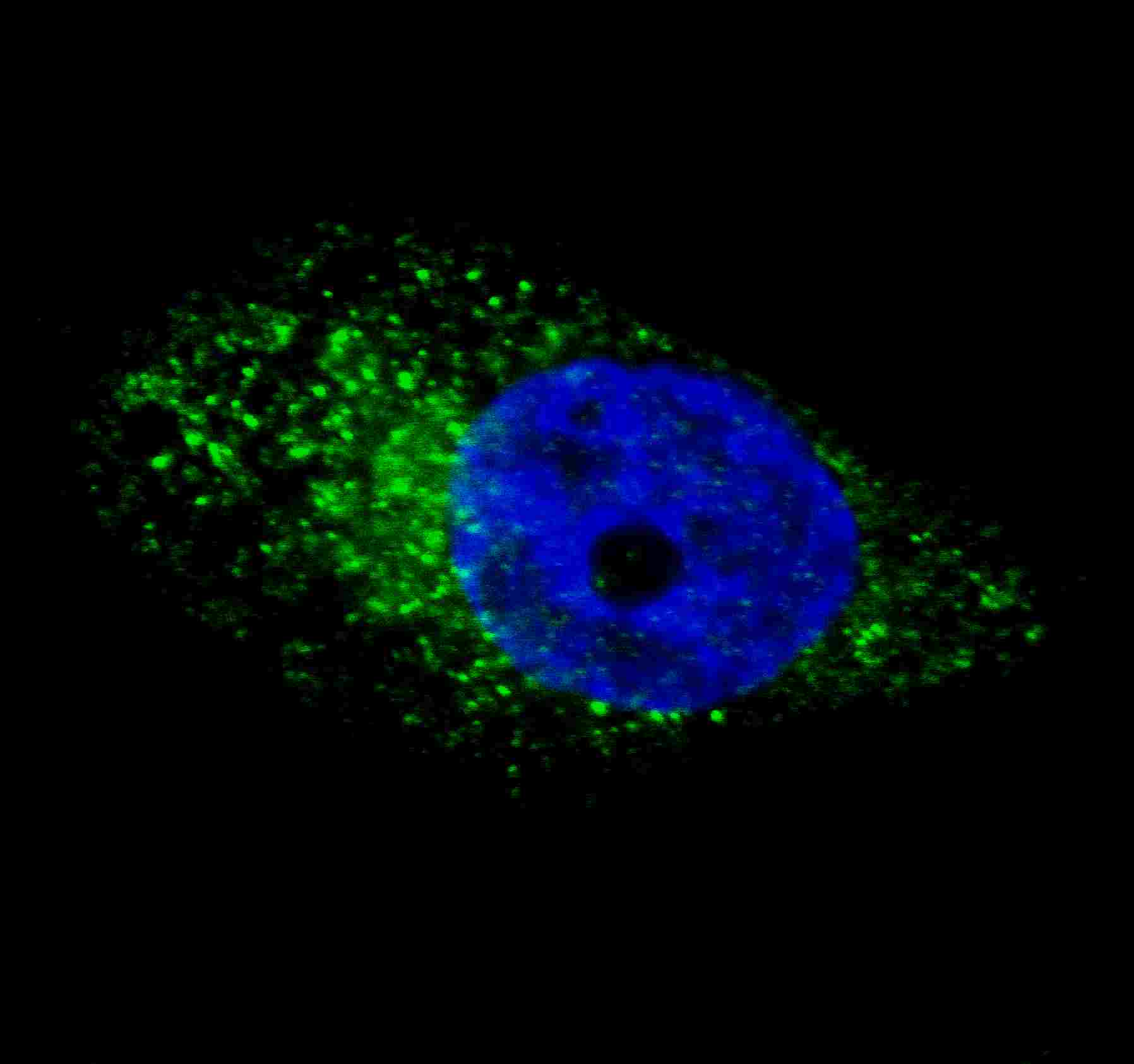
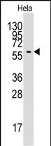
| WB, IF, E |
|---|---|
| Primary Accession | Q676U5 |
| Reactivity | Human |
| Host | Rabbit |
| Clonality | Polyclonal |
| Isotype | Rabbit IgG |
| Calculated MW | 68265 Da |
| Gene ID | 55054 |
|---|---|
| Other Names | Autophagy-related protein 16-1, APG16-like 1, ATG16L1, APG16L |
| Target/Specificity | This APG16L antibody is generated from rabbits immunized with a recombinant fragment protein from human APG16L. |
| Dilution | IF~~1:100 WB~~1:1000 |
| Format | Purified polyclonal antibody supplied in PBS with 0.09% (W/V) sodium azide. This antibody is prepared by Saturated Ammonium Sulfate (SAS) precipitation followed by dialysis against PBS. |
| Storage | Maintain refrigerated at 2-8°C for up to 2 weeks. For long term storage store at -20°C in small aliquots to prevent freeze-thaw cycles. |
| Precautions | ATG16L Antibody is for research use only and not for use in diagnostic or therapeutic procedures. |
| Name | ATG16L1 {ECO:0000303|PubMed:17200669, ECO:0000312|HGNC:HGNC:21498} |
|---|---|
| Function | Plays an essential role in both canonical and non-canonical autophagy: interacts with ATG12-ATG5 to mediate the lipidation to ATG8 family proteins (MAP1LC3A, MAP1LC3B, MAP1LC3C, GABARAPL1, GABARAPL2 and GABARAP) (PubMed:23376921, PubMed:23392225, PubMed:29317426, PubMed:30778222, PubMed:33909989, PubMed:24553140, PubMed:24954904, PubMed:27273576). Acts as a molecular hub, coordinating autophagy pathways via distinct domains that support either canonical or non- canonical signaling (PubMed:29317426, PubMed:30778222). During canonical autophagy, interacts with ATG12-ATG5 to mediate the conjugation of phosphatidylethanolamine (PE) to ATG8 proteins, to produce a membrane-bound activated form of ATG8 (PubMed:23376921, PubMed:23392225, PubMed:24553140, PubMed:24954904, PubMed:27273576). Thereby, controls the elongation of the nascent autophagosomal membrane (PubMed:23376921, PubMed:23392225, PubMed:24553140, PubMed:24954904, PubMed:27273576). Also involved in non-canonical autophagy, a parallel pathway involving conjugation of ATG8 proteins to single membranes at endolysosomal compartments, probably by catalyzing conjugation of phosphatidylserine (PS) to ATG8 (PubMed:33909989). Non-canonical autophagy plays a key role in epithelial cells to limit lethal infection by influenza A (IAV) virus (By similarity). Regulates mitochondrial antiviral signaling (MAVS)-dependent type I interferon (IFN-I) production (PubMed:22749352, PubMed:25645662). Negatively regulates NOD1- and NOD2-driven inflammatory cytokine response (PubMed:24238340). Instead, promotes an autophagy-dependent antibacterial pathway together with NOD1 or NOD2 (PubMed:20637199). Plays a role in regulating morphology and function of Paneth cell (PubMed:18849966). |
| Cellular Location | Cytoplasm. Preautophagosomal structure membrane; Peripheral membrane protein. Endosome membrane; Peripheral membrane protein. Lysosome membrane; Peripheral membrane protein. Note=Recruited to omegasomes membranes by WIPI2 (By similarity). Omegasomes are endoplasmic reticulum connected strutures at the origin of preautophagosomal structures (By similarity) Localized to preautophagosomal structure (PAS) where it is involved in the membrane targeting of ATG5 (By similarity). Localizes also to discrete punctae along the ciliary axoneme (By similarity). Upon activation of non-canonical autophagy, recruited to single-membrane endolysosomal compartments (PubMed:29317426) {ECO:0000250|UniProtKB:Q8C0J2, ECO:0000269|PubMed:29317426} |

Provided below are standard protocols that you may find useful for product applications.
Background
Macroautophagy is the major inducible pathway for the general turnover of cytoplasmic constituents in eukaryotic cells, it is also responsible for the degradation of active cytoplasmic enzymes and organelles during nutrient starvation. Macroautophagy involves the formation of double-membrane bound autophagosomes which enclose the cytoplasmic constituent targeted for degradation in a membrane bound structure, which then fuse with the lysosome (or vacuole) releasing a single-membrane bound autophagic bodies which are then degraded within the lysosome (or vacuole). The APG12-APG5-APG16L complex is esential for the elongation of autophagic isolation membranes. This complex initially associates in uniform distribution with small vesicle membranes. During membrane elongation, the complex partitions, with a great concentration building on the outer side of the isolation membrane. Upon completion of the formation of the autophagosome, the APG12-APG5-APG16L dissociates from the membrane.
References
References for protein:
1.Baehrecke EH. Nat Rev Mol Cell Biol. 6(6):505-10. (2005)
2.Lum JJ, et al. Nat Rev Mol Cell Biol. 6(6):439-48. (2005)
3.Greenberg JT. Dev Cell. 8(6):799-801. (2005)
4.Levine B. Cell. 120(2):159-62. (2005)
5.Shintani T and Klionsky DJ. Science. 306(5698):990-5. (2004)
References for U251 cell line:
1. Westermark B.; Pontén J.; Hugosson R. (1973).” Determinants for the establishment of permanent tissue culture lines from human gliomas”. Acta Pathol Microbiol Scand A. 81:791-805. [PMID: 4359449].
2. Pontén, J.,Westermark B. (1978).” Properties of Human Malignant Glioma Cells in Vitro”. Medical Biology 56: 184-193.[PMID: 359950].
3. Geng Y.;Kohli L.; Klocke B.J.; Roth K.A.(2010). “Chloroquine-induced autophagic vacuole accumulation and cell death in glioma cells is p53 independent”. Neuro Oncol. 12(5): 473–481.[ PMID: 20406898].
If you have used an Abcepta product and would like to share how it has performed, please click on the "Submit Review" button and provide the requested information. Our staff will examine and post your review and contact you if needed.
If you have any additional inquiries please email technical services at tech@abcepta.com.

Ordering Information
Other Products
Shipping Information
