


 Foundational characteristics of cancer include proliferation, angiogenesis, migration, evasion of apoptosis, and cellular immortality. Find key markers for these cellular processes and antibodies to detect them.
 The SUMOplot™ Analysis Program predicts and scores sumoylation sites in your protein. SUMOylation is a post-translational modification involved in various cellular processes, such as nuclear-cytosolic transport, transcriptional regulation, apoptosis, protein stability, response to stress, and progression through the cell cycle.
 The Autophagy Receptor Motif Plotter predicts and scores autophagy receptor binding sites in your protein. Identifying proteins connected to this pathway is critical to understanding the role of autophagy in physiological as well as pathological processes such as development, differentiation, neurodegenerative diseases, stress, infection, and cancer.
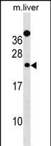
Mouse Derl2 Antibody (C-term)
Affinity Purified Rabbit Polyclonal Antibody (Pab)
- SPECIFICATION
- CITATIONS
- PROTOCOLS
- BACKGROUND

Application
| WB, E |
|---|---|
| Primary Accession | Q8BNI4 |
| Other Accession | NP_291040.1 |
| Reactivity | Mouse |
| Host | Rabbit |
| Clonality | Polyclonal |
| Isotype | Rabbit IgG |
| Calculated MW | 27640 Da |
| Antigen Region | 191-219 aa |
| Gene ID | 116891 |
|---|---|
| Other Names | Derlin-2, Degradation in endoplasmic reticulum protein 2, Der1-like protein 2, F-LANa, Derl2, Der2, Flana |
| Target/Specificity | This Mouse Derl2 antibody is generated from rabbits immunized with a KLH conjugated synthetic peptide between 191-219 amino acids from the C-terminal region of mouse Derl2. |
| Dilution | WB~~1:1000 |
| Format | Purified polyclonal antibody supplied in PBS with 0.09% (W/V) sodium azide. This antibody is purified through a protein A column, followed by peptide affinity purification. |
| Storage | Maintain refrigerated at 2-8°C for up to 2 weeks. For long term storage store at -20°C in small aliquots to prevent freeze-thaw cycles. |
| Precautions | Mouse Derl2 Antibody (C-term) is for research use only and not for use in diagnostic or therapeutic procedures. |
| Name | Derl2 {ECO:0000312|MGI:MGI:2151483} |
|---|---|
| Function | Functional component of endoplasmic reticulum-associated degradation (ERAD) for misfolded lumenal glycoproteins, but not that of misfolded nonglycoproteins. May act by forming a channel that allows the retrotranslocation of misfolded glycoproteins into the cytosol where they are ubiquitinated and degraded by the proteasome. May mediate the interaction between VCP and misfolded glycoproteins. May also be involved in endoplasmic reticulum stress-induced pre-emptive quality control, a mechanism that selectively attenuates the translocation of newly synthesized proteins into the endoplasmic reticulum and reroutes them to the cytosol for proteasomal degradation. |
| Cellular Location | Endoplasmic reticulum membrane {ECO:0000250|UniProtKB:Q9GZP9}; Multi-pass membrane protein {ECO:0000250|UniProtKB:Q9GZP9} |
| Tissue Location | Widely expressed, with lowest levels in brain and heart. |

Thousands of laboratories across the world have published research that depended on the performance of antibodies from Abcepta to advance their research. Check out links to articles that cite our products in major peer-reviewed journals, organized by research category.
info@abcepta.com, and receive a free "I Love Antibodies" mug.

Provided below are standard protocols that you may find useful for product applications.
Background
Functional component of endoplasmic reticulum-associated degradation (ERAD) for misfolded lumenal glycoproteins, but not that of misfolded nonglycoproteins. May act by forming a channel that allows the retrotranslocation of misfolded glycoproteins into the cytosol where they are ubiquitinated and degraded by the proteasome. May mediate the interaction between VCP and the degradation substrate (By similarity).
References
Schaheen, B., et al. J. Cell. Sci. 122 (PT 13), 2228-2239 (2009) :
Oda, Y., et al. J. Cell Biol. 172(3):383-393(2006)
Lilley, B.N., et al. Proc. Natl. Acad. Sci. U.S.A. 102(40):14296-14301(2005)
Kaput, J., et al. Physiol. Genomics 18(3):316-324(2004)
Lilley, B.N., et al. Nature 429(6994):834-840(2004)
If you have used an Abcepta product and would like to share how it has performed, please click on the "Submit Review" button and provide the requested information. Our staff will examine and post your review and contact you if needed.
If you have any additional inquiries please email technical services at tech@abcepta.com.
Ordering Information
Other Products
Shipping Information
