


 Foundational characteristics of cancer include proliferation, angiogenesis, migration, evasion of apoptosis, and cellular immortality. Find key markers for these cellular processes and antibodies to detect them.
 The SUMOplot™ Analysis Program predicts and scores sumoylation sites in your protein. SUMOylation is a post-translational modification involved in various cellular processes, such as nuclear-cytosolic transport, transcriptional regulation, apoptosis, protein stability, response to stress, and progression through the cell cycle.
 The Autophagy Receptor Motif Plotter predicts and scores autophagy receptor binding sites in your protein. Identifying proteins connected to this pathway is critical to understanding the role of autophagy in physiological as well as pathological processes such as development, differentiation, neurodegenerative diseases, stress, infection, and cancer.
GUCA1B Antibody(N-term)
Affinity Purified Rabbit Polyclonal Antibody (Pab)
- SPECIFICATION
- CITATIONS
- PROTOCOLS
- BACKGROUND

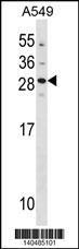
Application
| WB, E |
|---|---|
| Primary Accession | Q9UMX6 |
| Other Accession | NP_002089.4 |
| Reactivity | Human |
| Host | Rabbit |
| Clonality | Polyclonal |
| Isotype | Rabbit IgG |
| Calculated MW | 23420 Da |
| Antigen Region | 29-57 aa |
| Gene ID | 2979 |
|---|---|
| Other Names | Guanylyl cyclase-activating protein 2, GCAP 2, Guanylate cyclase activator 1B, GUCA1B, GCAP2 |
| Target/Specificity | This GUCA1B antibody is generated from rabbits immunized with a KLH conjugated synthetic peptide between 29-57 amino acids from the N-terminal region of human GUCA1B. |
| Dilution | WB~~1:1000 |
| Format | Purified polyclonal antibody supplied in PBS with 0.09% (W/V) sodium azide. This antibody is purified through a protein A column, followed by peptide affinity purification. |
| Storage | Maintain refrigerated at 2-8°C for up to 2 weeks. For long term storage store at -20°C in small aliquots to prevent freeze-thaw cycles. |
| Precautions | GUCA1B Antibody(N-term) is for research use only and not for use in diagnostic or therapeutic procedures. |
| Name | GUCA1B |
|---|---|
| Synonyms | GCAP2 |
| Function | Stimulates two retinal guanylyl cyclases (GCs) GUCY2D and GUCY2F when free calcium ions concentration is low, and inhibits GUCY2D and GUCY2F when free calcium ions concentration is elevated (By similarity). This Ca(2+)-sensitive regulation of GCs is a key event in recovery of the dark state of rod photoreceptors following light exposure (By similarity). May be involved in cone photoreceptor response and recovery of response in bright light (By similarity). |
| Cellular Location | Cell membrane {ECO:0000250|UniProtKB:P51177}; Lipid-anchor {ECO:0000250|UniProtKB:P51177}. Photoreceptor inner segment. Cell projection, cilium, photoreceptor outer segment Note=Subcellular location is not affected by light or dark conditions {ECO:0000250|UniProtKB:Q8VBV8} |
| Tissue Location | In the retina, it is expressed in cone and rod photoreceptor cells. |

Thousands of laboratories across the world have published research that depended on the performance of antibodies from Abcepta to advance their research. Check out links to articles that cite our products in major peer-reviewed journals, organized by research category.
info@abcepta.com, and receive a free "I Love Antibodies" mug.

Provided below are standard protocols that you may find useful for product applications.
Background
The protein encoded by this gene is a calcium-binding protein that activates photoreceptor guanylate cyclases. This gene may have arisen due to a gene duplication event since there is a highly similar gene clustered with it on chromosome 6. Mutations in this gene can cause a form of retinitis pigmentosa. [provided by RefSeq].
References
Sato, M., et al. Graefes Arch. Clin. Exp. Ophthalmol. 243(3):235-242(2005)
Nishiguchi, K.M., et al. Invest. Ophthalmol. Vis. Sci. 45(11):3863-3870(2004)
Mungall, A.J., et al. Nature 425(6960):805-811(2003)
Wistow, G., et al. Mol. Vis. 8, 196-204 (2002) :
Zhang, Q.J., et al. Yi Chuan 24(1):19-21(2002)
If you have used an Abcepta product and would like to share how it has performed, please click on the "Submit Review" button and provide the requested information. Our staff will examine and post your review and contact you if needed.
If you have any additional inquiries please email technical services at tech@abcepta.com.
Ordering Information
Shipping Information
