


 Foundational characteristics of cancer include proliferation, angiogenesis, migration, evasion of apoptosis, and cellular immortality. Find key markers for these cellular processes and antibodies to detect them.
 The SUMOplot™ Analysis Program predicts and scores sumoylation sites in your protein. SUMOylation is a post-translational modification involved in various cellular processes, such as nuclear-cytosolic transport, transcriptional regulation, apoptosis, protein stability, response to stress, and progression through the cell cycle.
 The Autophagy Receptor Motif Plotter predicts and scores autophagy receptor binding sites in your protein. Identifying proteins connected to this pathway is critical to understanding the role of autophagy in physiological as well as pathological processes such as development, differentiation, neurodegenerative diseases, stress, infection, and cancer.
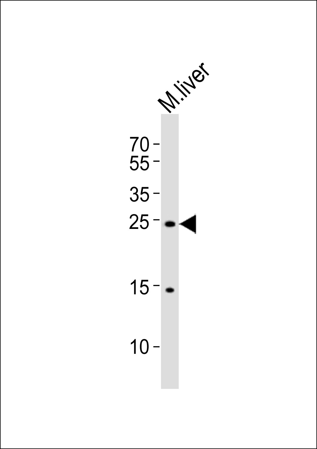
PDCD6 Antibody (Center)
Purified Rabbit Polyclonal Antibody (Pab)
- SPECIFICATION
- CITATIONS
- PROTOCOLS
- BACKGROUND

Application
| WB, E |
|---|---|
| Primary Accession | O75340 |
| Other Accession | P12815 |
| Reactivity | Mouse |
| Host | Rabbit |
| Clonality | Polyclonal |
| Isotype | Rabbit IgG |
| Calculated MW | 21868 Da |
| Gene ID | 10016 |
|---|---|
| Other Names | Programmed cell death protein 6, Apoptosis-linked gene 2 protein, Probable calcium-binding protein ALG-2, PDCD6, ALG2 |
| Target/Specificity | This PDCD6 antibody is generated from a rabbit immunized with a KLH conjugated synthetic peptide between 103-137 amino acids from the Central region of human PDCD6. |
| Dilution | WB~~1:1000 |
| Format | Purified polyclonal antibody supplied in PBS with 0.09% (W/V) sodium azide. This antibody is purified through a protein A column, followed by peptide affinity purification. |
| Storage | Maintain refrigerated at 2-8°C for up to 2 weeks. For long term storage store at -20°C in small aliquots to prevent freeze-thaw cycles. |
| Precautions | PDCD6 Antibody (Center) is for research use only and not for use in diagnostic or therapeutic procedures. |
| Name | PDCD6 |
|---|---|
| Synonyms | ALG2 {ECO:0000250|UniProtKB:P12815} |
| Function | Calcium sensor that plays a key role in processes such as endoplasmic reticulum (ER)-Golgi vesicular transport, endosomal biogenesis or membrane repair. Acts as an adapter that bridges unrelated proteins or stabilizes weak protein-protein complexes in response to calcium: calcium-binding triggers exposure of apolar surface, promoting interaction with different sets of proteins thanks to 3 different hydrophobic pockets, leading to translocation to membranes (PubMed:20691033, PubMed:25667979). Involved in ER-Golgi transport by promoting the association between PDCD6IP and TSG101, thereby bridging together the ESCRT-III and ESCRT-I complexes (PubMed:19520058). Together with PEF1, acts as a calcium-dependent adapter for the BCR(KLHL12) complex, a complex involved in ER-Golgi transport by regulating the size of COPII coats (PubMed:27716508). In response to cytosolic calcium increase, the heterodimer formed with PEF1 interacts with, and bridges together the BCR(KLHL12) complex and SEC31 (SEC31A or SEC31B), promoting monoubiquitination of SEC31 and subsequent collagen export, which is required for neural crest specification (PubMed:27716508). Involved in the regulation of the distribution and function of MCOLN1 in the endosomal pathway (PubMed:19864416). Promotes localization and polymerization of TFG at endoplasmic reticulum exit site (PubMed:27813252). Required for T-cell receptor-, Fas-, and glucocorticoid-induced apoptosis (By similarity). May mediate Ca(2+)-regulated signals along the death pathway: interaction with DAPK1 can accelerate apoptotic cell death by increasing caspase-3 activity (PubMed:16132846). Its role in apoptosis may however be indirect, as suggested by knockout experiments (By similarity). May inhibit KDR/VEGFR2-dependent angiogenesis; the function involves inhibition of VEGF-induced phosphorylation of the Akt signaling pathway (PubMed:21893193). In case of infection by HIV-1 virus, indirectly inhibits HIV-1 production by affecting viral Gag expression and distribution (PubMed:27784779). |
| Cellular Location | Endoplasmic reticulum membrane; Peripheral membrane protein. Cytoplasmic vesicle, COPII-coated vesicle membrane. Cytoplasm. Nucleus. Endosome Note=Interaction with RBM22 induces relocalization from the cytoplasm to the nucleus (PubMed:17045351). Translocated from the cytoplasm to the nucleus after heat shock cell treatment. Accumulates in cytoplasmic vesicle-like organelles after heat shock treatment, which may represent stress granules (PubMed:21122810). In response to calcium increase, relocates from cytoplasm to COPII vesicle coat (PubMed:27716508) Localizes to endoplasmic reticulum exit site (ERES) (PubMed:27813252) |

Thousands of laboratories across the world have published research that depended on the performance of antibodies from Abcepta to advance their research. Check out links to articles that cite our products in major peer-reviewed journals, organized by research category.
info@abcepta.com, and receive a free "I Love Antibodies" mug.

Provided below are standard protocols that you may find useful for product applications.
Background
Calcium-binding protein required for T-cell receptor-, Fas-, and glucocorticoid-induced cell death. May mediate Ca(2+)- regulated signals along the death pathway (By similarity). Calcium-dependent adapter necessary for the association between PDCD6IP and TSG101. Interaction with DAPK1 can accelerate apoptotic cell death by increasing caspase-3 activity. May inhibit KDR/VEGFR2-dependent angiogenesis; the function involves inhibition of VEGF-induced phosphoprylation of the Akt signaling pathway. Seems to play a role in the regulation of the distribution and function of MCOLN1 in the endosomal pathway. Isoform 2 has a lower Ca(2+) affinity than isoform 1. Isoform 1 and, to a lesser extend, isoform 2, can stabilize SHISA5.
References
Ganjei J.K.,et al.Submitted (NOV-1997) to the EMBL/GenBank/DDBJ databases.
Urcelay E.,et al.Submitted (MAY-1996) to the EMBL/GenBank/DDBJ databases.
Ota T.,et al.Nat. Genet. 36:40-45(2004).
Kalnine N.,et al.Submitted (OCT-2004) to the EMBL/GenBank/DDBJ databases.
Schmutz J.,et al.Nature 431:268-274(2004).
If you have used an Abcepta product and would like to share how it has performed, please click on the "Submit Review" button and provide the requested information. Our staff will examine and post your review and contact you if needed.
If you have any additional inquiries please email technical services at tech@abcepta.com.

Ordering Information
Other Products
Shipping Information
