


 Foundational characteristics of cancer include proliferation, angiogenesis, migration, evasion of apoptosis, and cellular immortality. Find key markers for these cellular processes and antibodies to detect them.
 The SUMOplot™ Analysis Program predicts and scores sumoylation sites in your protein. SUMOylation is a post-translational modification involved in various cellular processes, such as nuclear-cytosolic transport, transcriptional regulation, apoptosis, protein stability, response to stress, and progression through the cell cycle.
 The Autophagy Receptor Motif Plotter predicts and scores autophagy receptor binding sites in your protein. Identifying proteins connected to this pathway is critical to understanding the role of autophagy in physiological as well as pathological processes such as development, differentiation, neurodegenerative diseases, stress, infection, and cancer.
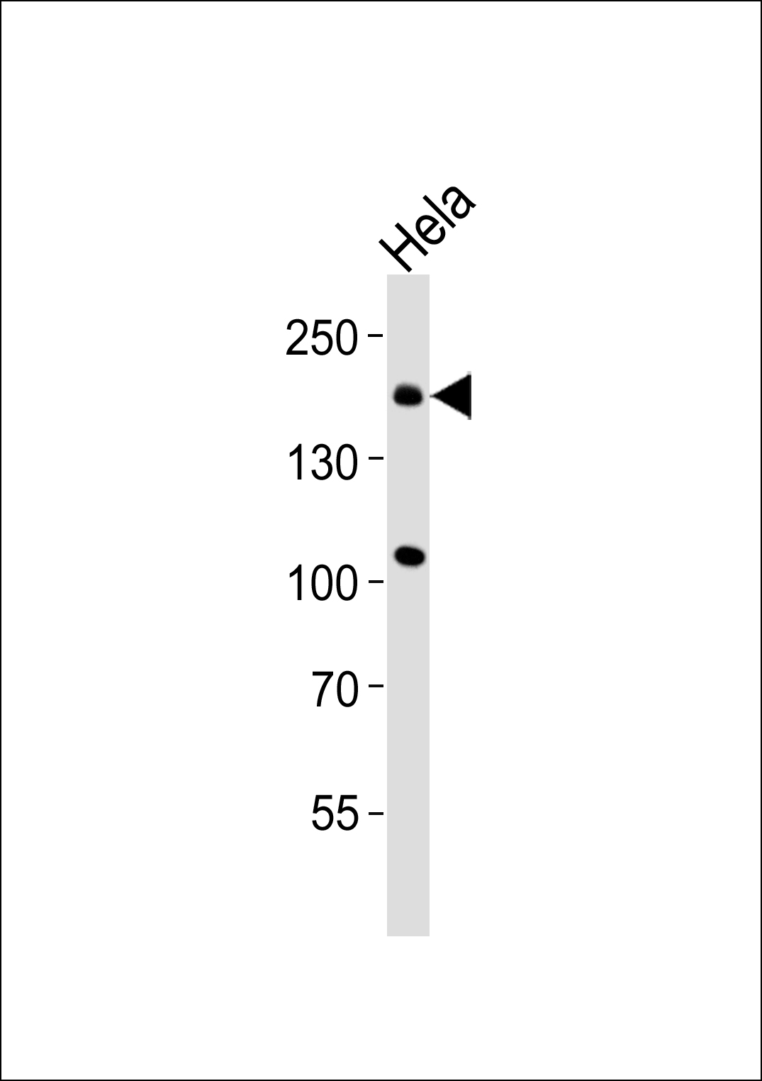
CLASP1 Antibody
Purified Rabbit Polyclonal Antibody (Pab)

- SPECIFICATION
- CITATIONS
- PROTOCOLS
- BACKGROUND

Application
| WB, IHC |
|---|---|
| Primary Accession | Q7Z460 |
| Reactivity | Human, Mouse |
| Host | Rabbit |
| Clonality | Polyclonal |
| Calculated MW | 169,162,165,163 KDa |
| Antigen Region | 1190-1218 aa |
| Gene ID | 23332 |
|---|---|
| Other Names | CLIP-associating protein 1, Cytoplasmic linker-associated protein 1, Multiple asters homolog 1, Protein Orbit homolog 1, hOrbit1, CLASP1, KIAA0622, MAST1 |
| Dilution | WB~~ 1:1000 IHC~~1:50~100 |
| Format | Rabbit IgG in phosphate buffered saline (without Mg2+ and Ca2+), pH 7.4, 150mM NaCl, 0.09% (W/V) sodium azide and 50% glycerol. |
| Storage Conditions | -20℃ |
| Name | CLASP1 |
|---|---|
| Synonyms | KIAA0622, MAST1 |
| Function | Microtubule plus-end tracking protein that promotes the stabilization of dynamic microtubules. Involved in the nucleation of noncentrosomal microtubules originating from the trans-Golgi network (TGN). Required for the polarization of the cytoplasmic microtubule arrays in migrating cells towards the leading edge of the cell. May act at the cell cortex to enhance the frequency of rescue of depolymerizing microtubules by attaching their plus-ends to cortical platforms composed of ERC1 and PHLDB2. This cortical microtubule stabilizing activity is regulated at least in part by phosphatidylinositol 3-kinase signaling. Also performs a similar stabilizing function at the kinetochore which is essential for the bipolar alignment of chromosomes on the mitotic spindle. |
| Cellular Location | Cytoplasm, cytoskeleton. Cytoplasm, cytoskeleton, microtubule organizing center, centrosome. Chromosome, centromere, kinetochore Cytoplasm, cytoskeleton, spindle. Golgi apparatus, trans-Golgi network. Note=Localizes to microtubule plus ends. Localizes to centrosomes, kinetochores and the mitotic spindle from prometaphase Subsequently localizes to the spindle midzone from anaphase and to the midbody from telophase. In migrating cells localizes to the plus ends of microtubules within the cell body and to the entire microtubule lattice within the lamella. Localizes to the cell cortex and this requires ERC1 and PHLDB2 |

Thousands of laboratories across the world have published research that depended on the performance of antibodies from Abcepta to advance their research. Check out links to articles that cite our products in major peer-reviewed journals, organized by research category.
info@abcepta.com, and receive a free "I Love Antibodies" mug.

Provided below are standard protocols that you may find useful for product applications.
Background
Microtubule plus-end tracking protein that promotes the stabilization of dynamic microtubules. Involved in the nucleation of noncentrosomal microtubules originating from the trans-Golgi network (TGN). Required for the polarization of the cytoplasmic microtubule arrays in migrating cells towards the leading edge of the cell. May act at the cell cortex to enhance the frequency of rescue of depolymerizing microtubules by attaching their plus-ends to cortical platforms composed of ERC1 and PHLDB2. This cortical microtubule stabilizing activity is regulated at least in part by phosphatidylinositol 3-kinase signaling. Also performs a similar stabilizing function at the kinetochore which is essential for the bipolar alignment of chromosomes on the mitotic spindle.
References
Maiato H.,et al.Submitted (FEB-2001) to the EMBL/GenBank/DDBJ databases.
Hillier L.W.,et al.Nature 434:724-731(2005).
Akhmanova A.,et al.Cell 104:923-935(2001).
Bechtel S.,et al.BMC Genomics 8:399-399(2007).
Ishikawa K.,et al.DNA Res. 5:169-176(1998).
If you have used an Abcepta product and would like to share how it has performed, please click on the "Submit Review" button and provide the requested information. Our staff will examine and post your review and contact you if needed.
If you have any additional inquiries please email technical services at tech@abcepta.com.
Ordering Information
Other Products
Shipping Information
