


 Foundational characteristics of cancer include proliferation, angiogenesis, migration, evasion of apoptosis, and cellular immortality. Find key markers for these cellular processes and antibodies to detect them.
 The SUMOplot™ Analysis Program predicts and scores sumoylation sites in your protein. SUMOylation is a post-translational modification involved in various cellular processes, such as nuclear-cytosolic transport, transcriptional regulation, apoptosis, protein stability, response to stress, and progression through the cell cycle.
 The Autophagy Receptor Motif Plotter predicts and scores autophagy receptor binding sites in your protein. Identifying proteins connected to this pathway is critical to understanding the role of autophagy in physiological as well as pathological processes such as development, differentiation, neurodegenerative diseases, stress, infection, and cancer.
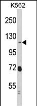
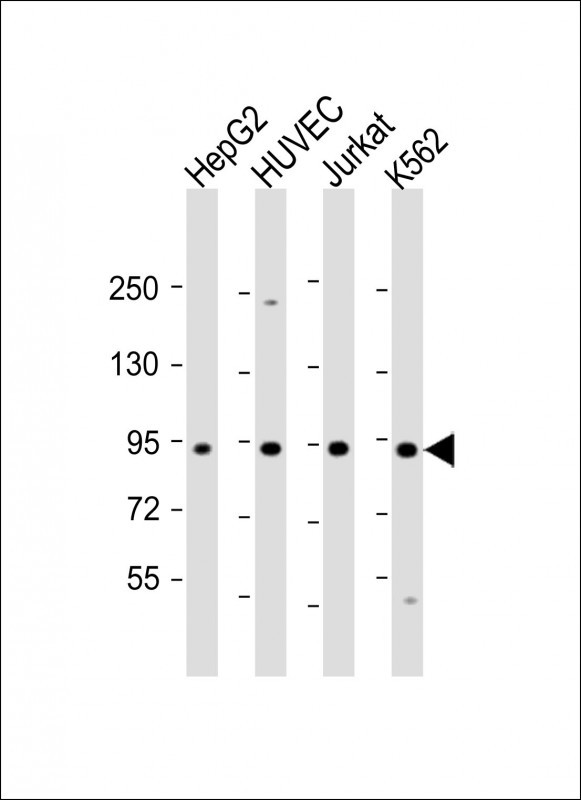
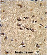
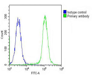
RASIP1 Antibody (Center)
Purified Rabbit Polyclonal Antibody (Pab)
- SPECIFICATION
- CITATIONS: 1
- PROTOCOLS
- BACKGROUND

Application
| WB, IHC-P, FC, E |
|---|---|
| Primary Accession | Q5U651 |
| Reactivity | Human |
| Host | Rabbit |
| Clonality | Polyclonal |
| Isotype | Rabbit IgG |
| Calculated MW | 103457 Da |
| Antigen Region | 651-678 aa |
| Gene ID | 54922 |
|---|---|
| Other Names | Ras-interacting protein 1, Rain, RASIP1 |
| Target/Specificity | This RASIP1 antibody is generated from rabbits immunized with a KLH conjugated synthetic peptide between 651-678 amino acids from the Central region of human RASIP1. |
| Dilution | WB~~1:2000 IHC-P~~1:50~100 FC~~1:25 |
| Format | Purified polyclonal antibody supplied in PBS with 0.09% (W/V) sodium azide. This antibody is purified through a protein A column, followed by peptide affinity purification. |
| Storage | Maintain refrigerated at 2-8°C for up to 2 weeks. For long term storage store at -20°C in small aliquots to prevent freeze-thaw cycles. |
| Precautions | RASIP1 Antibody (Center) is for research use only and not for use in diagnostic or therapeutic procedures. |
| Name | RASIP1 |
|---|---|
| Function | Required for the proper formation of vascular structures that develop via both vasculogenesis and angiogenesis. Acts as a critical and vascular-specific regulator of GTPase signaling, cell architecture, and adhesion, which is essential for endothelial cell morphogenesis and blood vessel tubulogenesis. Regulates the activity of Rho GTPases in part by recruiting ARHGAP29 and suppressing RhoA signaling and dampening ROCK and MYH9 activities in endothelial cells (By similarity). May act as effector for Golgi-bound HRAS and other Ras- like proteins. May promote HRAS-mediated transformation. Negative regulator of amino acid starvation-induced autophagy. |
| Cellular Location | Cytoplasm, perinuclear region. Golgi apparatus, Golgi stack. Note=Associated with perinuclear vesicles. Is recruited to Golgi stacks by activated HRAS |
| Tissue Location | Highly expressed in heart. Detected at lower levels in placenta and pancreas. |
Research Areas
Citations ( 0 )

Application Protocols
Provided below are standard protocols that you may find useful for product applications.
Background
RASIP1 may act as effector for Golgi-bound HRAS and other Ras-like proteins. May promote HRAS-mediated transformation.
References
Mitin,N.Y.,et.al., J. Biol. Chem. 279 (21), 22353-22361 (2004)
Abcepta welcomes feedback from its customers.
If you have used an Abcepta product and would like to share how it has performed, please click on the "Submit Review" button and provide the requested information. Our staff will examine and post your review and contact you if needed.
If you have any additional inquiries please email technical services at tech@abcepta.com.
$ 365.00
$ 140.00
Cat# AP6870c
Ordering Information
United States
AlbaniaAustraliaAustriaBelgiumBosnia & HerzegovinaBrazilBulgariaCanadaCentral AmericaChinaCroatiaCyprusCzech RepublicDenmarkEstoniaFinlandFranceGermanyGreeceHong KongHungaryIcelandIndiaIndonesiaIrelandIsraelItalyJapanLatviaLithuaniaLuxembourgMacedoniaMalaysiaMaltaNetherlandsNew ZealandNorwayPakistanPolandPortugalRomaniaSerbiaSingaporeSlovakiaSloveniaSouth AfricaSouth KoreaSpainSwedenSwitzerlandTaiwanTurkeyUnited KingdomUnited StatesVietnamWorldwideOthers
USA Headquarters
(888) 735-7227 / (858) 622-0099 or (858) 875-1900
Shipping Information
Domestic orders (in stock items)
Shipped out the same day. Orders placed after 1 PM (PST) will ship out the next business day.
International orders
Contact your local distributors
