


 Foundational characteristics of cancer include proliferation, angiogenesis, migration, evasion of apoptosis, and cellular immortality. Find key markers for these cellular processes and antibodies to detect them.
 The SUMOplot™ Analysis Program predicts and scores sumoylation sites in your protein. SUMOylation is a post-translational modification involved in various cellular processes, such as nuclear-cytosolic transport, transcriptional regulation, apoptosis, protein stability, response to stress, and progression through the cell cycle.
 The Autophagy Receptor Motif Plotter predicts and scores autophagy receptor binding sites in your protein. Identifying proteins connected to this pathway is critical to understanding the role of autophagy in physiological as well as pathological processes such as development, differentiation, neurodegenerative diseases, stress, infection, and cancer.
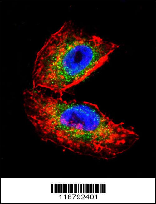
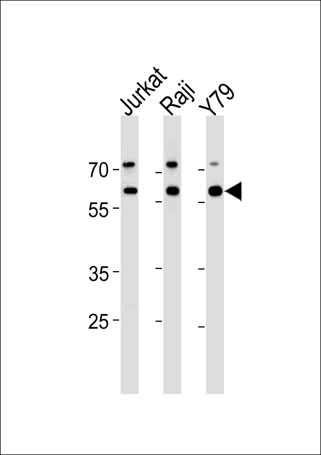
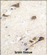
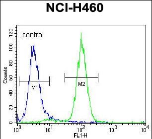
ACHE Antibody (N-term)
Purified Rabbit Polyclonal Antibody (Pab)
- SPECIFICATION
- CITATIONS
- PROTOCOLS
- BACKGROUND

Application
| WB, IF, FC, IHC-P, E |
|---|---|
| Primary Accession | P22303 |
| Reactivity | Human |
| Predicted | Guinea Pig |
| Host | Rabbit |
| Clonality | Polyclonal |
| Isotype | Rabbit IgG |
| Calculated MW | 67796 Da |
| Antigen Region | 147-175 aa |
| Gene ID | 43 |
|---|---|
| Other Names | Acetylcholinesterase, AChE, ACHE |
| Target/Specificity | This ACHE antibody is generated from rabbits immunized with a KLH conjugated synthetic peptide between 147-175 amino acids from the N-terminal region of human ACHE. |
| Dilution | IF~~1:10~50 WB~~1:1000 IHC-P~~1:10~50 FC~~1:10~50 |
| Format | Purified polyclonal antibody supplied in PBS with 0.09% (W/V) sodium azide. This antibody is prepared by Saturated Ammonium Sulfate (SAS) precipitation followed by dialysis against PBS. |
| Storage | Maintain refrigerated at 2-8°C for up to 2 weeks. For long term storage store at -20°C in small aliquots to prevent freeze-thaw cycles. |
| Precautions | ACHE Antibody (N-term) is for research use only and not for use in diagnostic or therapeutic procedures. |
| Name | ACHE (HGNC:108) |
|---|---|
| Function | Hydrolyzes rapidly the acetylcholine neurotransmitter released into the synaptic cleft allowing to terminate the signal transduction at the neuromuscular junction. Role in neuronal apoptosis. |
| Cellular Location | Synapse. Secreted. Cell membrane; Peripheral membrane protein [Isoform H]: Cell membrane; Lipid- anchor, GPI-anchor; Extracellular side |
| Tissue Location | Isoform H is highly expressed in erythrocytes. |

Thousands of laboratories across the world have published research that depended on the performance of antibodies from Abcepta to advance their research. Check out links to articles that cite our products in major peer-reviewed journals, organized by research category.
info@abcepta.com, and receive a free "I Love Antibodies" mug.

Provided below are standard protocols that you may find useful for product applications.
Background
Acetylcholinesterase hydrolyzes the neurotransmitter, acetylcholine at neuromuscular junctions and brain cholinergic synapses, and thus terminates signal transmission. The Protein is also found on the red blood cell membranes, where it constitutes the Yt blood group antigen. Acetylcholinesterase exists in multiple molecular forms which possess similar catalytic properties, but differ in their oligomeric assembly and mode of cell attachment to the cell surface. The major form of acetylcholinesterase found in brain, muscle and other tissues is the hydrophilic species, which forms disulfide-linked oligomers with collagenous, or lipid-containing structural subunits.
References
Liang,D., FEBS J. 276 (1), 94-108 (2009)
Scacchi,R., Am. J. Med. Genet. B Neuropsychiatr. Genet. (2008)
If you have used an Abcepta product and would like to share how it has performed, please click on the "Submit Review" button and provide the requested information. Our staff will examine and post your review and contact you if needed.
If you have any additional inquiries please email technical services at tech@abcepta.com.
Ordering Information
Other Products
Shipping Information
