


 Foundational characteristics of cancer include proliferation, angiogenesis, migration, evasion of apoptosis, and cellular immortality. Find key markers for these cellular processes and antibodies to detect them.
 The SUMOplot™ Analysis Program predicts and scores sumoylation sites in your protein. SUMOylation is a post-translational modification involved in various cellular processes, such as nuclear-cytosolic transport, transcriptional regulation, apoptosis, protein stability, response to stress, and progression through the cell cycle.
 The Autophagy Receptor Motif Plotter predicts and scores autophagy receptor binding sites in your protein. Identifying proteins connected to this pathway is critical to understanding the role of autophagy in physiological as well as pathological processes such as development, differentiation, neurodegenerative diseases, stress, infection, and cancer.
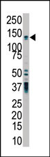
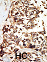
PKD2 Antibody (C-term)
Purified Rabbit Polyclonal Antibody (Pab)
- SPECIFICATION
- CITATIONS
- PROTOCOLS
- BACKGROUND

Application
| WB, IHC-P, E |
|---|---|
| Primary Accession | Q13563 |
| Reactivity | Human |
| Host | Rabbit |
| Clonality | Polyclonal |
| Isotype | Rabbit IgG |
| Calculated MW | 109691 Da |
| Antigen Region | 937-968 aa |
| Gene ID | 5311 |
|---|---|
| Other Names | Polycystin-2, Autosomal dominant polycystic kidney disease type II protein, Polycystic kidney disease 2 protein, Polycystwin, R48321, PKD2 |
| Target/Specificity | This PKD2 antibody is generated from rabbits immunized with a KLH conjugated synthetic peptide between 937-968 amino acids from the C-terminal region of human PKD2. |
| Dilution | WB~~1:1000 IHC-P~~1:50~100 |
| Format | Purified polyclonal antibody supplied in PBS with 0.09% (W/V) sodium azide. This antibody is prepared by Saturated Ammonium Sulfate (SAS) precipitation followed by dialysis against PBS. |
| Storage | Maintain refrigerated at 2-8°C for up to 2 weeks. For long term storage store at -20°C in small aliquots to prevent freeze-thaw cycles. |
| Precautions | PKD2 Antibody (C-term) is for research use only and not for use in diagnostic or therapeutic procedures. |
| Name | PKD2 (HGNC:9009) |
|---|---|
| Function | Component of a heteromeric calcium-permeable ion channel formed by PKD1 and PKD2 that is activated by interaction between PKD1 and a Wnt family member, such as WNT3A and WNT9B (PubMed:27214281). Can also form a functional, homotetrameric ion channel (PubMed:29899465). Functions as a cation channel involved in fluid-flow mechanosensation by the primary cilium in renal epithelium (PubMed:18695040). Functions as outward-rectifying K(+) channel, but is also permeable to Ca(2+), and to a much lesser degree also to Na(+) (PubMed:11854751, PubMed:15692563, PubMed:27071085, PubMed:27991905). May contribute to the release of Ca(2+) stores from the endoplasmic reticulum (PubMed:11854751, PubMed:20881056). Together with TRPV4, forms mechano- and thermosensitive channels in cilium (PubMed:18695040). PKD1 and PKD2 may function through a common signaling pathway that is necessary to maintain the normal, differentiated state of renal tubule cells. Acts as a regulator of cilium length, together with PKD1. The dynamic control of cilium length is essential in the regulation of mechanotransductive signaling. The cilium length response creates a negative feedback loop whereby fluid shear-mediated deflection of the primary cilium, which decreases intracellular cAMP, leads to cilium shortening and thus decreases flow-induced signaling. Also involved in left-right axis specification via its role in sensing nodal flow; forms a complex with PKD1L1 in cilia to facilitate flow detection in left- right patterning. Detection of asymmetric nodal flow gives rise to a Ca(2+) signal that is required for normal, asymmetric expression of genes involved in the specification of body left-right laterality (By similarity). |
| Cellular Location | Cell projection, cilium membrane; Multi-pass membrane protein. Endoplasmic reticulum membrane; Multi-pass membrane protein. Cell membrane; Multi-pass membrane protein. Basolateral cell membrane. Cytoplasmic vesicle membrane. Golgi apparatus {ECO:0000250|UniProtKB:O35245}. Vesicle Secreted, extracellular exosome Note=PKD2 localization to the plasma and ciliary membranes requires PKD1. PKD1:PKD2 interaction is required to reach the Golgi apparatus form endoplasmic reticulum and then traffic to the cilia (By similarity). Retained in the endoplasmic reticulum by interaction with PACS1 and PACS2 (PubMed:15692563). Detected on kidney tubule basolateral membranes and basal cytoplasmic vesicles (PubMed:10770959) Cell surface and cilium localization requires GANAB (PubMed:27259053) Detected on migrasomes and on extracellular exosomes in urine (PubMed:21406692). {ECO:0000250|UniProtKB:O35245, ECO:0000269|PubMed:15692563, ECO:0000269|PubMed:21406692, ECO:0000269|PubMed:27259053} |
| Tissue Location | Detected in fetal and adult kidney (PubMed:10770959). Detected at the thick ascending limb of the loop of Henle, at distal tubules, including the distal convoluted tubule and cortical collecting tubules, with weak staining of the collecting duct (PubMed:10770959). Detected on placenta syncytiotrophoblasts (at protein level) (PubMed:26269590). Strongly expressed in ovary, fetal and adult kidney, testis, and small intestine. Not detected in peripheral leukocytes. |

Thousands of laboratories across the world have published research that depended on the performance of antibodies from Abcepta to advance their research. Check out links to articles that cite our products in major peer-reviewed journals, organized by research category.
info@abcepta.com, and receive a free "I Love Antibodies" mug.

Provided below are standard protocols that you may find useful for product applications.
Background
Involved in fluid-flow mechanosensation by the primary cilium in renal epithelium. PKD1 and PKD2 may function through a common signaling pathway that is necessary for normal tubulogenesis. Acts as a regulator of cilium length, together with PKD1. The dynamic control of cilium length is essential in the regulation of mechanotransductive signaling. The cilium length response creates a negative feedback loop whereby fluid shear-mediated deflection of the primary cilium, which decreases intracellular cAMP, leads to cilium shortening and thus decreases flow-induced signaling. Functions as a calcium permeable cation channel.
If you have used an Abcepta product and would like to share how it has performed, please click on the "Submit Review" button and provide the requested information. Our staff will examine and post your review and contact you if needed.
If you have any additional inquiries please email technical services at tech@abcepta.com.

Ordering Information
Other Products
Shipping Information
