


 Foundational characteristics of cancer include proliferation, angiogenesis, migration, evasion of apoptosis, and cellular immortality. Find key markers for these cellular processes and antibodies to detect them.
 The SUMOplot™ Analysis Program predicts and scores sumoylation sites in your protein. SUMOylation is a post-translational modification involved in various cellular processes, such as nuclear-cytosolic transport, transcriptional regulation, apoptosis, protein stability, response to stress, and progression through the cell cycle.
 The Autophagy Receptor Motif Plotter predicts and scores autophagy receptor binding sites in your protein. Identifying proteins connected to this pathway is critical to understanding the role of autophagy in physiological as well as pathological processes such as development, differentiation, neurodegenerative diseases, stress, infection, and cancer.
MARK2 (EMK) Antibody (C-term)
Purified Rabbit Polyclonal Antibody (Pab)

- SPECIFICATION
- CITATIONS: 1
- PROTOCOLS
- BACKGROUND

Application
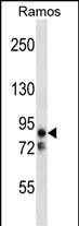
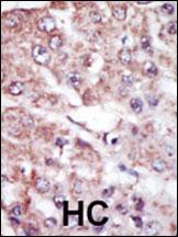
| WB, IHC-P, E |
|---|---|
| Primary Accession | Q7KZI7 |
| Other Accession | O08679, Q05512, Q15524 |
| Reactivity | Human, Mouse |
| Predicted | Rat |
| Host | Rabbit |
| Clonality | Polyclonal |
| Isotype | Rabbit IgG |
| Calculated MW | 87911 Da |
| Antigen Region | 600-630 aa |
| Gene ID | 2011 |
|---|---|
| Other Names | Serine/threonine-protein kinase MARK2, ELKL motif kinase 1, EMK-1, MAP/microtubule affinity-regulating kinase 2, PAR1 homolog, PAR1 homolog b, Par-1b, Par1b, MARK2 {ECO:0000312|EMBL:AAH087712}, EMK1 |
| Target/Specificity | This MARK2 (EMK) antibody is generated from rabbits immunized with a KLH conjugated synthetic peptide between 600-630 amino acids from the C-terminal region of human MARK2 (EMK). |
| Dilution | WB~~1:1000 IHC-P~~1:50~100 |
| Format | Purified polyclonal antibody supplied in PBS with 0.09% (W/V) sodium azide. This antibody is purified through a protein A column, followed by peptide affinity purification. |
| Storage | Maintain refrigerated at 2-8°C for up to 2 weeks. For long term storage store at -20°C in small aliquots to prevent freeze-thaw cycles. |
| Precautions | MARK2 (EMK) Antibody (C-term) is for research use only and not for use in diagnostic or therapeutic procedures. |
| Name | MARK2 {ECO:0000312|EMBL:AAH08771.2} |
|---|---|
| Synonyms | EMK1 |
| Function | Serine/threonine-protein kinase (PubMed:23666762). Involved in cell polarity and microtubule dynamics regulation. Phosphorylates CRTC2/TORC2, DCX, HDAC7, KIF13B, MAP2, MAP4 and RAB11FIP2. Phosphorylates the microtubule-associated protein MAPT/TAU (PubMed:23666762). Plays a key role in cell polarity by phosphorylating the microtubule-associated proteins MAP2, MAP4 and MAPT/TAU at KXGS motifs, causing detachment from microtubules, and their disassembly. Regulates epithelial cell polarity by phosphorylating RAB11FIP2. Involved in the regulation of neuronal migration through its dual activities in regulating cellular polarity and microtubule dynamics, possibly by phosphorylating and regulating DCX. Regulates axogenesis by phosphorylating KIF13B, promoting interaction between KIF13B and 14-3-3 and inhibiting microtubule-dependent accumulation of KIF13B. Also required for neurite outgrowth and establishment of neuronal polarity. Regulates localization and activity of some histone deacetylases by mediating phosphorylation of HDAC7, promoting subsequent interaction between HDAC7 and 14-3-3 and export from the nucleus. Also acts as a positive regulator of the Wnt signaling pathway, probably by mediating phosphorylation of dishevelled proteins (DVL1, DVL2 and/or DVL3). Modulates the developmental decision to build a columnar versus a hepatic epithelial cell apparently by promoting a switch from a direct to a transcytotic mode of apical protein delivery. Essential for the asymmetric development of membrane domains of polarized epithelial cells. |
| Cellular Location | Cell membrane; Peripheral membrane protein. Cytoplasm. Lateral cell membrane. Cytoplasm, cytoskeleton. Cell projection, dendrite. Cytoplasm. Note=Phosphorylation at Thr-596 by PRKCZ/aPKC and subsequent interaction with 14-3-3 protein YWHAZ promotes relocation from the cell membrane to the cytoplasm |
| Tissue Location | High levels of expression in heart, brain, skeletal muscle and pancreas, lower levels observed in lung, liver and kidney |

Provided below are standard protocols that you may find useful for product applications.
Background
Protein kinases are enzymes that transfer a phosphate group from a phosphate donor, generally the g phosphate of ATP, onto an acceptor amino acid in a substrate protein. By this basic mechanism, protein kinases mediate most of the signal transduction in eukaryotic cells, regulating cellular metabolism, transcription, cell cycle progression, cytoskeletal rearrangement and cell movement, apoptosis, and differentiation. With more than 500 gene products, the protein kinase family is one of the largest families of proteins in eukaryotes. The family has been classified in 8 major groups based on sequence comparison of their tyrosine (PTK) or serine/threonine (STK) kinase catalytic domains.
References
Blume-Jensen P, et al. Nature 2001. 411: 355.
Cantrell D, J. Cell Sci. 2001. 114: 1439.
Jhiang S Oncogene 2000. 19: 5590.
Manning G, et al. Science 2002. 298: 1912.
Moller, D, et al. Am. J. Physiol. 1994. 266: C351-C359.
Robertson, S. et al. Trends Genet. 2000. 16: 368.
Robinson D, et al. Oncogene 2000. 19: 5548.
Van der Ven, P, et al. Hum. Molec. Genet. 1993. 2: 1889.
Vanhaesebroeck, B, et al. Biochem. J. 2000. 346: 561.
Van Weering D, et al. Recent Results Cancer Res. 1998. 154: 271.
If you have used an Abcepta product and would like to share how it has performed, please click on the "Submit Review" button and provide the requested information. Our staff will examine and post your review and contact you if needed.
If you have any additional inquiries please email technical services at tech@abcepta.com.

Ordering Information
Other Products
Shipping Information
