


 Foundational characteristics of cancer include proliferation, angiogenesis, migration, evasion of apoptosis, and cellular immortality. Find key markers for these cellular processes and antibodies to detect them.
 The SUMOplot™ Analysis Program predicts and scores sumoylation sites in your protein. SUMOylation is a post-translational modification involved in various cellular processes, such as nuclear-cytosolic transport, transcriptional regulation, apoptosis, protein stability, response to stress, and progression through the cell cycle.
 The Autophagy Receptor Motif Plotter predicts and scores autophagy receptor binding sites in your protein. Identifying proteins connected to this pathway is critical to understanding the role of autophagy in physiological as well as pathological processes such as development, differentiation, neurodegenerative diseases, stress, infection, and cancer.
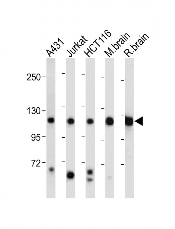
PI3KCA Antibody (Center)
Purified Rabbit Polyclonal Antibody (Pab)
- SPECIFICATION
- CITATIONS: 1
- PROTOCOLS
- BACKGROUND

Application
| WB, E |
|---|---|
| Primary Accession | P42336 |
| Other Accession | Q14CW1 |
| Reactivity | Human, Mouse, Rat |
| Host | Rabbit |
| Clonality | Polyclonal |
| Isotype | Rabbit IgG |
| Calculated MW | 124284 Da |
| Antigen Region | 504-533 aa |
| Gene ID | 5290 |
|---|---|
| Other Names | Phosphatidylinositol 4, 5-bisphosphate 3-kinase catalytic subunit alpha isoform, PI3-kinase subunit alpha, PI3K-alpha, PI3Kalpha, PtdIns-3-kinase subunit alpha, Phosphatidylinositol 4, 5-bisphosphate 3-kinase 110 kDa catalytic subunit alpha, PtdIns-3-kinase subunit p110-alpha, p110alpha, Phosphoinositide-3-kinase catalytic alpha polypeptide, Serine/threonine protein kinase PIK3CA, PIK3CA |
| Target/Specificity | This PI3KCA antibody is generated from rabbits immunized with a KLH conjugated synthetic peptide between 504-533 amino acids from the Central region of human PI3KCA. |
| Dilution | WB~~1:2000 |
| Format | Purified polyclonal antibody supplied in PBS with 0.09% (W/V) sodium azide. This antibody is prepared by Saturated Ammonium Sulfate (SAS) precipitation followed by dialysis against PBS. |
| Storage | Maintain refrigerated at 2-8°C for up to 2 weeks. For long term storage store at -20°C in small aliquots to prevent freeze-thaw cycles. |
| Precautions | PI3KCA Antibody (Center) is for research use only and not for use in diagnostic or therapeutic procedures. |
| Name | PIK3CA |
|---|---|
| Function | Phosphoinositide-3-kinase (PI3K) phosphorylates phosphatidylinositol (PI) and its phosphorylated derivatives at position 3 of the inositol ring to produce 3-phosphoinositides (PubMed:15135396, PubMed:23936502, PubMed:28676499). Uses ATP and PtdIns(4,5)P2 (phosphatidylinositol 4,5-bisphosphate) to generate phosphatidylinositol 3,4,5-trisphosphate (PIP3) (PubMed:15135396, PubMed:28676499). PIP3 plays a key role by recruiting PH domain- containing proteins to the membrane, including AKT1 and PDPK1, activating signaling cascades involved in cell growth, survival, proliferation, motility and morphology. Participates in cellular signaling in response to various growth factors. Involved in the activation of AKT1 upon stimulation by receptor tyrosine kinases ligands such as EGF, insulin, IGF1, VEGFA and PDGF. Involved in signaling via insulin-receptor substrate (IRS) proteins. Essential in endothelial cell migration during vascular development through VEGFA signaling, possibly by regulating RhoA activity. Required for lymphatic vasculature development, possibly by binding to RAS and by activation by EGF and FGF2, but not by PDGF. Regulates invadopodia formation through the PDPK1-AKT1 pathway. Participates in cardiomyogenesis in embryonic stem cells through a AKT1 pathway. Participates in vasculogenesis in embryonic stem cells through PDK1 and protein kinase C pathway. In addition to its lipid kinase activity, it displays a serine-protein kinase activity that results in the autophosphorylation of the p85alpha regulatory subunit as well as phosphorylation of other proteins such as 4EBP1, H-Ras, the IL-3 beta c receptor and possibly others (PubMed:23936502, PubMed:28676499). Plays a role in the positive regulation of phagocytosis and pinocytosis (By similarity). |

Provided below are standard protocols that you may find useful for product applications.
Background
Phosphatidylinositol 3-kinase is composed of an 85 kDa regulatory subunit and a 110 kDa catalytic subunit. The protein encoded by this gene represents the catalytic subunit, which uses ATP to phosphorylate PtdIns, PtdIns4P and PtdIns(4,5)P2. This gene has been found to be oncogenic and has been implicated in cervical cancers.
References
Tiwari, S. Nat Immunol. August; 10(8): 907?17 (2009).
Ballou, L.M., et al., J. Biol. Chem. 278(26):23472-23479 (2003).
Singh, B., et al., Genes Dev. 16(8):984-993 (2002).
Shayesteh, L., et al., Nat. Genet. 21(1):99-102 (1999).
Volinia, S., et al., Genomics 24(3):472-477 (1994).
Hiles, I.D., et al., Cell 70(3):419-429 (1992).
If you have used an Abcepta product and would like to share how it has performed, please click on the "Submit Review" button and provide the requested information. Our staff will examine and post your review and contact you if needed.
If you have any additional inquiries please email technical services at tech@abcepta.com.
Ordering Information
Other Products
Shipping Information
