


 Foundational characteristics of cancer include proliferation, angiogenesis, migration, evasion of apoptosis, and cellular immortality. Find key markers for these cellular processes and antibodies to detect them.
 The SUMOplot™ Analysis Program predicts and scores sumoylation sites in your protein. SUMOylation is a post-translational modification involved in various cellular processes, such as nuclear-cytosolic transport, transcriptional regulation, apoptosis, protein stability, response to stress, and progression through the cell cycle.
 The Autophagy Receptor Motif Plotter predicts and scores autophagy receptor binding sites in your protein. Identifying proteins connected to this pathway is critical to understanding the role of autophagy in physiological as well as pathological processes such as development, differentiation, neurodegenerative diseases, stress, infection, and cancer.
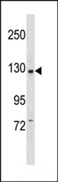
PI3KCB Antibody (C-term)
Purified Rabbit Polyclonal Antibody (Pab)
- SPECIFICATION
- CITATIONS: 1
- PROTOCOLS
- BACKGROUND

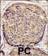
Application
| IHC-P, WB, E |
|---|---|
| Primary Accession | P42338 |
| Reactivity | Human |
| Host | Rabbit |
| Clonality | Polyclonal |
| Isotype | Rabbit IgG |
| Calculated MW | 122762 Da |
| Antigen Region | 713-744 aa |
| Gene ID | 5291 |
|---|---|
| Other Names | Phosphatidylinositol 4, 5-bisphosphate 3-kinase catalytic subunit beta isoform, PI3-kinase subunit beta, PI3K-beta, PI3Kbeta, PtdIns-3-kinase subunit beta, Phosphatidylinositol 4, 5-bisphosphate 3-kinase 110 kDa catalytic subunit beta, PtdIns-3-kinase subunit p110-beta, p110beta, PIK3CB, PIK3C1 |
| Target/Specificity | This PI3KCB antibody is generated from rabbits immunized with a KLH conjugated synthetic peptide between 713-744 amino acids from the C-terminal region of human PI3KCB. |
| Dilution | WB~~1:1000 IHC-P~~1:10~50 |
| Format | Purified polyclonal antibody supplied in PBS with 0.09% (W/V) sodium azide. This antibody is prepared by Saturated Ammonium Sulfate (SAS) precipitation followed by dialysis against PBS. |
| Storage | Maintain refrigerated at 2-8°C for up to 2 weeks. For long term storage store at -20°C in small aliquots to prevent freeze-thaw cycles. |
| Precautions | PI3KCB Antibody (C-term) is for research use only and not for use in diagnostic or therapeutic procedures. |
| Name | PIK3CB |
|---|---|
| Synonyms | PIK3C1 |
| Function | Phosphoinositide-3-kinase (PI3K) phosphorylates phosphatidylinositol derivatives at position 3 of the inositol ring to produce 3-phosphoinositides (PubMed:15135396). Uses ATP and PtdIns(4,5)P2 (phosphatidylinositol 4,5-bisphosphate) to generate phosphatidylinositol 3,4,5-trisphosphate (PIP3) (PubMed:15135396). PIP3 plays a key role by recruiting PH domain-containing proteins to the membrane, including AKT1 and PDPK1, activating signaling cascades involved in cell growth, survival, proliferation, motility and morphology. Involved in the activation of AKT1 upon stimulation by G- protein coupled receptors (GPCRs) ligands such as CXCL12, sphingosine 1-phosphate, and lysophosphatidic acid. May also act downstream receptor tyrosine kinases. Required in different signaling pathways for stable platelet adhesion and aggregation. Plays a role in platelet activation signaling triggered by GPCRs, alpha-IIb/beta-3 integrins (ITGA2B/ ITGB3) and ITAM (immunoreceptor tyrosine-based activation motif)-bearing receptors such as GP6. Regulates the strength of adhesion of ITGA2B/ ITGB3 activated receptors necessary for the cellular transmission of contractile forces. Required for platelet aggregation induced by F2 (thrombin) and thromboxane A2 (TXA2). Has a role in cell survival. May have a role in cell migration. Involved in the early stage of autophagosome formation. Modulates the intracellular level of PtdIns3P (phosphatidylinositol 3-phosphate) and activates PIK3C3 kinase activity. May act as a scaffold, independently of its lipid kinase activity to positively regulate autophagy. May have a role in insulin signaling as scaffolding protein in which the lipid kinase activity is not required. May have a kinase-independent function in regulating cell proliferation and in clathrin-mediated endocytosis. Mediator of oncogenic signal in cell lines lacking PTEN. The lipid kinase activity is necessary for its role in oncogenic transformation. Required for the growth of ERBB2 and RAS driven tumors. Has also a protein kinase activity showing autophosphorylation (PubMed:12502714). |
| Cellular Location | Cytoplasm. Nucleus. Note=Interaction with PIK3R2 is required for nuclear localization and export |
| Tissue Location | Expressed ubiquitously. |

Provided below are standard protocols that you may find useful for product applications.
Background
Protein kinases are enzymes that transfer a phosphate group from a phosphate donor, generally the g phosphate of ATP, onto an acceptor amino acid in a substrate protein. By this basic mechanism, protein kinases mediate most of the signal transduction in eukaryotic cells, regulating cellular metabolism, transcription, cell cycle progression, cytoskeletal rearrangement and cell movement, apoptosis, and differentiation. With more than 500 gene products, the protein kinase family is one of the largest families of proteins in eukaryotes. The family has been classified in 8 major groups based on sequence comparison of their tyrosine (PTK) or serine/threonine (STK) kinase catalytic domains.
References
Brock, C., et al., J. Cell Biol. 160(1):89-99 (2003).
Kossila, M., et al., Diabetes Care 26(1):179-182 (2003).
Yart, A., et al., J. Biol. Chem. 277(24):21167-21178 (2002).
Ueki, K., et al., Proc. Natl. Acad. Sci. U.S.A. 99(1):419-424 (2002).
Sotsios, Y., et al., J. Immunol. 163(11):5954-5963 (1999).
If you have used an Abcepta product and would like to share how it has performed, please click on the "Submit Review" button and provide the requested information. Our staff will examine and post your review and contact you if needed.
If you have any additional inquiries please email technical services at tech@abcepta.com.
Ordering Information
Other Products
Shipping Information
