


 Foundational characteristics of cancer include proliferation, angiogenesis, migration, evasion of apoptosis, and cellular immortality. Find key markers for these cellular processes and antibodies to detect them.
 The SUMOplot™ Analysis Program predicts and scores sumoylation sites in your protein. SUMOylation is a post-translational modification involved in various cellular processes, such as nuclear-cytosolic transport, transcriptional regulation, apoptosis, protein stability, response to stress, and progression through the cell cycle.
 The Autophagy Receptor Motif Plotter predicts and scores autophagy receptor binding sites in your protein. Identifying proteins connected to this pathway is critical to understanding the role of autophagy in physiological as well as pathological processes such as development, differentiation, neurodegenerative diseases, stress, infection, and cancer.
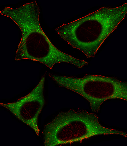
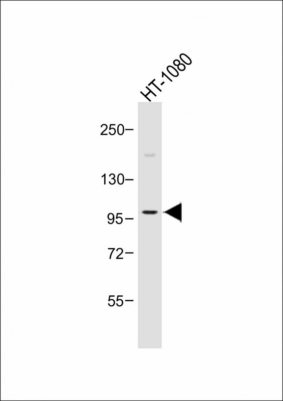
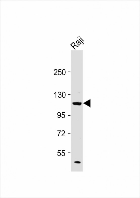
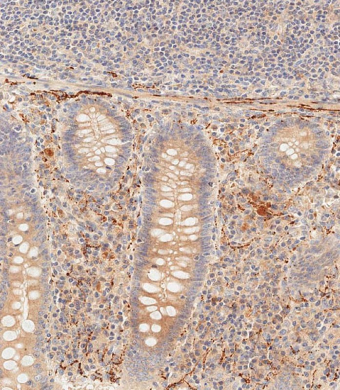
PI3KCD Antibody (C-term)
Purified Rabbit Polyclonal Antibody (Pab)
- SPECIFICATION
- CITATIONS
- PROTOCOLS
- BACKGROUND

Application
| IF, WB, IHC-P-Leica, E |
|---|---|
| Primary Accession | O00329 |
| Reactivity | Human |
| Host | Rabbit |
| Clonality | Polyclonal |
| Isotype | Rabbit IgG |
| Calculated MW | 119479 Da |
| Antigen Region | 687-717 aa |
| Gene ID | 5293 |
|---|---|
| Other Names | Phosphatidylinositol 4, 5-bisphosphate 3-kinase catalytic subunit delta isoform, PI3-kinase subunit delta, PI3K-delta, PI3Kdelta, PtdIns-3-kinase subunit delta, Phosphatidylinositol 4, 5-bisphosphate 3-kinase 110 kDa catalytic subunit delta, PtdIns-3-kinase subunit p110-delta, p110delta, PIK3CD |
| Target/Specificity | This PI3KCD antibody is generated from rabbits immunized with a KLH conjugated synthetic peptide between 687-717 amino acids from the C-terminal region of human PI3KCD. |
| Dilution | IF~~1:10~50 WB~~1:2000 IHC-P-Leica~~1:500 |
| Format | Purified polyclonal antibody supplied in PBS with 0.09% (W/V) sodium azide. This antibody is purified through a protein A column, followed by peptide affinity purification. |
| Storage | Maintain refrigerated at 2-8°C for up to 2 weeks. For long term storage store at -20°C in small aliquots to prevent freeze-thaw cycles. |
| Precautions | PI3KCD Antibody (C-term) is for research use only and not for use in diagnostic or therapeutic procedures. |
| Name | PIK3CD |
|---|---|
| Function | Phosphoinositide-3-kinase (PI3K) phosphorylates phosphatidylinositol (PI) and its phosphorylated derivatives at position 3 of the inositol ring to produce 3-phosphoinositides (PubMed:9235916). Uses ATP and PtdIns(4,5)P2 (phosphatidylinositol 4,5- bisphosphate) to generate phosphatidylinositol 3,4,5-trisphosphate (PIP3) (PubMed:15135396). PIP3 plays a key role by recruiting PH domain-containing proteins to the membrane, including AKT1 and PDPK1, activating signaling cascades involved in cell growth, survival, proliferation, motility and morphology. Mediates immune responses. Plays a role in B-cell development, proliferation, migration, and function. Required for B-cell receptor (BCR) signaling. Mediates B-cell proliferation response to anti-IgM, anti-CD40 and IL4 stimulation. Promotes cytokine production in response to TLR4 and TLR9. Required for antibody class switch mediated by TLR9. Involved in the antigen presentation function of B-cells. Involved in B-cell chemotaxis in response to CXCL13 and sphingosine 1-phosphate (S1P). Required for proliferation, signaling and cytokine production of naive, effector and memory T-cells. Required for T-cell receptor (TCR) signaling. Mediates TCR signaling events at the immune synapse. Activation by TCR leads to antigen-dependent memory T-cell migration and retention to antigenic tissues. Together with PIK3CG participates in T-cell development. Contributes to T-helper cell expansion and differentiation. Required for T-cell migration mediated by homing receptors SELL/CD62L, CCR7 and S1PR1 and antigen dependent recruitment of T-cells. Together with PIK3CG is involved in natural killer (NK) cell development and migration towards the sites of inflammation. Participates in NK cell receptor activation. Plays a role in NK cell maturation and cytokine production. Together with PIK3CG is involved in neutrophil chemotaxis and extravasation. Together with PIK3CG participates in neutrophil respiratory burst. Plays important roles in mast-cell development and mast cell mediated allergic response. Involved in stem cell factor (SCF)-mediated proliferation, adhesion and migration. Required for allergen-IgE-induced degranulation and cytokine release. The lipid kinase activity is required for its biological function. Isoform 2 may be involved in stabilizing total RAS levels, resulting in increased ERK phosphorylation and increased PI3K activity. |
| Cellular Location | Cytoplasm. |
| Tissue Location | In humans, the highest levels of expression are seen in peripheral blood mononuclear cells, spleen, and thymus, and low levels of expression in testes, uterus, colon, and small intestine but not in other tissues examined including prostate, heart, brain, and liver (PubMed:9235916). Isoform 2 is expressed in normal thymus, lung and spleen tissues, and is detected at low levels in normal lysates from colon and ovarian biopsies, at elevated levels in lysates from colorectal tumors and is abundantly expressed in some ovarian tumors (at protein level). Both isoform 1 and isoform 2 are widely expressed Isoform 1 is expressed predominantly in leukocytes |

Thousands of laboratories across the world have published research that depended on the performance of antibodies from Abcepta to advance their research. Check out links to articles that cite our products in major peer-reviewed journals, organized by research category.
info@abcepta.com, and receive a free "I Love Antibodies" mug.

Provided below are standard protocols that you may find useful for product applications.
Background
Protein kinases are enzymes that transfer a phosphate group from a phosphate donor, generally the g phosphate of ATP, onto an acceptor amino acid in a substrate protein. By this basic mechanism, protein kinases mediate most of the signal transduction in eukaryotic cells, regulating cellular metabolism, transcription, cell cycle progression, cytoskeletal rearrangement and cell movement, apoptosis, and differentiation. With more than 500 gene products, the protein kinase family is one of the largest families of proteins in eukaryotes. The family has been classified in 8 major groups based on sequence comparison of their tyrosine (PTK) or serine/threonine (STK) kinase catalytic domains.
References
Vanhaesebroeck, B., et al., Proc. Natl. Acad. Sci. U.S.A. 94(9):4330-4335 (1997).
Chantry, D., et al., J. Biol. Chem. 272(31):19236-19241 (1997).
If you have used an Abcepta product and would like to share how it has performed, please click on the "Submit Review" button and provide the requested information. Our staff will examine and post your review and contact you if needed.
If you have any additional inquiries please email technical services at tech@abcepta.com.
Ordering Information
Other Products
Shipping Information
