


 Foundational characteristics of cancer include proliferation, angiogenesis, migration, evasion of apoptosis, and cellular immortality. Find key markers for these cellular processes and antibodies to detect them.
 The SUMOplot™ Analysis Program predicts and scores sumoylation sites in your protein. SUMOylation is a post-translational modification involved in various cellular processes, such as nuclear-cytosolic transport, transcriptional regulation, apoptosis, protein stability, response to stress, and progression through the cell cycle.
 The Autophagy Receptor Motif Plotter predicts and scores autophagy receptor binding sites in your protein. Identifying proteins connected to this pathway is critical to understanding the role of autophagy in physiological as well as pathological processes such as development, differentiation, neurodegenerative diseases, stress, infection, and cancer.
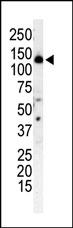
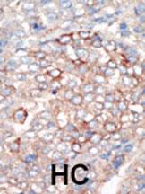
PI3KR4 Antibody (N-term)
Purified Rabbit Polyclonal Antibody (Pab)
- SPECIFICATION
- CITATIONS
- PROTOCOLS
- BACKGROUND

Application
| IHC-P, WB, E |
|---|---|
| Primary Accession | Q99570 |
| Other Accession | NP_055417 |
| Reactivity | Human |
| Host | Rabbit |
| Clonality | Polyclonal |
| Isotype | Rabbit IgG |
| Calculated MW | 153103 Da |
| Antigen Region | 53-83 aa |
| Gene ID | 30849 |
|---|---|
| Other Names | Phosphoinositide 3-kinase regulatory subunit 4, PI3-kinase regulatory subunit 4, PI3-kinase p150 subunit, Phosphoinositide 3-kinase adaptor protein, PIK3R4 |
| Target/Specificity | This PI3KR4 antibody is generated from rabbits immunized with a KLH conjugated synthetic peptide between 53-83 amino acids from the N-terminal region of human PI3KR4. |
| Dilution | WB~~1:1000 IHC-P~~1:50~100 |
| Format | Purified polyclonal antibody supplied in PBS with 0.09% (W/V) sodium azide. This antibody is prepared by Saturated Ammonium Sulfate (SAS) precipitation followed by dialysis against PBS. |
| Storage | Maintain refrigerated at 2-8°C for up to 2 weeks. For long term storage store at -20°C in small aliquots to prevent freeze-thaw cycles. |
| Precautions | PI3KR4 Antibody (N-term) is for research use only and not for use in diagnostic or therapeutic procedures. |
| Name | PIK3R4 |
|---|---|
| Synonyms | VPS15 {ECO:0000303|PubMed:23878393} |
| Function | Regulatory subunit of the PI3K complex that mediates formation of phosphatidylinositol 3-phosphate; different complex forms are believed to play a role in multiple membrane trafficking pathways: PI3KC3-C1 is involved in initiation of autophagosomes and PI3KC3-C2 in maturation of autophagosomes and endocytosis. Involved in regulation of degradative endocytic trafficking and cytokinesis, probably in the context of PI3KC3-C2 (PubMed:20643123). |
| Cellular Location | Late endosome. Cytoplasmic vesicle, autophagosome. Membrane; Lipid-anchor. Note=As component of the PI3K complex I localized to pre-autophagosome structures. As component of the PI3K complex II localized predominantly to endosomes. Localizes also to discrete punctae along the ciliary axoneme (By similarity) {ECO:0000250|UniProtKB:Q8VD65, ECO:0000305} |
| Tissue Location | Ubiquitously expressed. |

Thousands of laboratories across the world have published research that depended on the performance of antibodies from Abcepta to advance their research. Check out links to articles that cite our products in major peer-reviewed journals, organized by research category.
info@abcepta.com, and receive a free "I Love Antibodies" mug.

Provided below are standard protocols that you may find useful for product applications.
Background
Protein kinases are enzymes that transfer a phosphate group from a phosphate donor, generally the g phosphate of ATP, onto an acceptor amino acid in a substrate protein. By this basic mechanism, protein kinases mediate most of the signal transduction in eukaryotic cells, regulating cellular metabolism, transcription, cell cycle progression, cytoskeletal rearrangement and cell movement, apoptosis, and differentiation. With more than 500 gene products, the protein kinase family is one of the largest families of proteins in eukaryotes. The family has been classified in 8 major groups based on sequence comparison of their tyrosine (PTK) or serine/threonine (STK) kinase catalytic domains.
References
Panaretou, C., et al., J. Biol. Chem. 272(4):2477-2485 (1997).
If you have used an Abcepta product and would like to share how it has performed, please click on the "Submit Review" button and provide the requested information. Our staff will examine and post your review and contact you if needed.
If you have any additional inquiries please email technical services at tech@abcepta.com.
Ordering Information
Other Products
Shipping Information
