


 Foundational characteristics of cancer include proliferation, angiogenesis, migration, evasion of apoptosis, and cellular immortality. Find key markers for these cellular processes and antibodies to detect them.
 The SUMOplot™ Analysis Program predicts and scores sumoylation sites in your protein. SUMOylation is a post-translational modification involved in various cellular processes, such as nuclear-cytosolic transport, transcriptional regulation, apoptosis, protein stability, response to stress, and progression through the cell cycle.
 The Autophagy Receptor Motif Plotter predicts and scores autophagy receptor binding sites in your protein. Identifying proteins connected to this pathway is critical to understanding the role of autophagy in physiological as well as pathological processes such as development, differentiation, neurodegenerative diseases, stress, infection, and cancer.
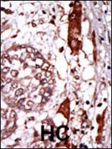
GNB2L1 Antibody (N-term)
Purified Rabbit Polyclonal Antibody (Pab)

- SPECIFICATION
- CITATIONS
- PROTOCOLS
- BACKGROUND

Application
| IHC-P, WB, E |
|---|---|
| Primary Accession | P63244 |
| Other Accession | P63245, P63246, P68040, Q4R7Y4, O42248, P63247, P63243, NP_006089, G1SJB4 |
| Reactivity | Human |
| Predicted | Zebrafish, Monkey, Bovine, Chicken, Mouse, Pig, Rabbit, Rat |
| Host | Rabbit |
| Clonality | Polyclonal |
| Isotype | Rabbit IgG |
| Calculated MW | 35077 Da |
| Antigen Region | 29-58 aa |
| Gene ID | 10399 |
|---|---|
| Other Names | Guanine nucleotide-binding protein subunit beta-2-like 1, Cell proliferation-inducing gene 21 protein, Guanine nucleotide-binding protein subunit beta-like protein 123, Human lung cancer oncogene 7 protein, HLC-7, Receptor for activated C kinase, Receptor of activated protein kinase C 1, RACK1, Guanine nucleotide-binding protein subunit beta-2-like 1, N-terminally processed, GNB2L1 |
| Target/Specificity | This GNB2L1 antibody is generated from rabbits immunized with a KLH conjugated synthetic peptide between 29-58 amino acids from the N-terminal region of human GNB2L1. |
| Dilution | WB~~1:1000 IHC-P~~1:50~100 |
| Format | Purified polyclonal antibody supplied in PBS with 0.09% (W/V) sodium azide. This antibody is prepared by Saturated Ammonium Sulfate (SAS) precipitation followed by dialysis against PBS. |
| Storage | Maintain refrigerated at 2-8°C for up to 2 weeks. For long term storage store at -20°C in small aliquots to prevent freeze-thaw cycles. |
| Precautions | GNB2L1 Antibody (N-term) is for research use only and not for use in diagnostic or therapeutic procedures. |
| Name | RACK1 (HGNC:4399) |
|---|---|
| Synonyms | GNB2L1 |
| Function | Scaffolding protein involved in the recruitment, assembly and/or regulation of a variety of signaling molecules. Interacts with a wide variety of proteins and plays a role in many cellular processes. Component of the 40S ribosomal subunit involved in translational repression (PubMed:23636399). Involved in the initiation of the ribosome quality control (RQC), a pathway that takes place when a ribosome has stalled during translation, by promoting ubiquitination of a subset of 40S ribosomal subunits (PubMed:28132843). Binds to and stabilizes activated protein kinase C (PKC), increasing PKC-mediated phosphorylation. May recruit activated PKC to the ribosome, leading to phosphorylation of EIF6. Inhibits the activity of SRC kinases including SRC, LCK and YES1. Inhibits cell growth by prolonging the G0/G1 phase of the cell cycle. Enhances phosphorylation of BMAL1 by PRKCA and inhibits transcriptional activity of the BMAL1-CLOCK heterodimer. Facilitates ligand-independent nuclear translocation of AR following PKC activation, represses AR transactivation activity and is required for phosphorylation of AR by SRC. Modulates IGF1R-dependent integrin signaling and promotes cell spreading and contact with the extracellular matrix. Involved in PKC-dependent translocation of ADAM12 to the cell membrane. Promotes the ubiquitination and proteasome- mediated degradation of proteins such as CLEC1B and HIF1A. Required for VANGL2 membrane localization, inhibits Wnt signaling, and regulates cellular polarization and oriented cell division during gastrulation. Required for PTK2/FAK1 phosphorylation and dephosphorylation. Regulates internalization of the muscarinic receptor CHRM2. Promotes apoptosis by increasing oligomerization of BAX and disrupting the interaction of BAX with the anti-apoptotic factor BCL2L. Inhibits TRPM6 channel activity. Regulates cell surface expression of some GPCRs such as TBXA2R. Plays a role in regulation of FLT1-mediated cell migration. Involved in the transport of ABCB4 from the Golgi to the apical bile canalicular membrane (PubMed:19674157). Promotes migration of breast carcinoma cells by binding to and activating RHOA (PubMed:20499158). Acts as an adapter for the dephosphorylation and inactivation of AKT1 by promoting recruitment of PP2A phosphatase to AKT1 (By similarity). |
| Cellular Location | Cell membrane; Peripheral membrane protein. Cytoplasm. Cytoplasm, perinuclear region. Nucleus. Perikaryon {ECO:0000250|UniProtKB:P68040}. Cell projection, dendrite {ECO:0000250|UniProtKB:P68040}. Cell projection, phagocytic cup. Note=Recruited to the plasma membrane through interaction with KRT1 which binds to membrane-bound ITGB1 (PubMed:17956333). Also associated with the membrane in oncogene- transformed cells (PubMed:11884618). PKC activation induces translocation from the perinuclear region to the cell periphery (PubMed:11279199). In the brain, detected mainly in cell bodies and dendrites with little expression in axonal fibers or nuclei (By similarity). Localized to phagocytic cups following infection by Y.pestis (PubMed:21347310). {ECO:0000250|UniProtKB:P68040, ECO:0000269|PubMed:11279199, ECO:0000269|PubMed:11884618, ECO:0000269|PubMed:17956333, ECO:0000269|PubMed:21347310} |
| Tissue Location | In the liver, expressed at higher levels in activated hepatic stellate cells than in hepatocytes or Kupffer cells Up-regulated in hepatocellular carcinomas and in the adjacent non-tumor liver tissue. |

Thousands of laboratories across the world have published research that depended on the performance of antibodies from Abcepta to advance their research. Check out links to articles that cite our products in major peer-reviewed journals, organized by research category.
info@abcepta.com, and receive a free "I Love Antibodies" mug.

Provided below are standard protocols that you may find useful for product applications.
Background
Protein kinases are enzymes that transfer a phosphate group from a phosphate donor, generally the g phosphate of ATP, onto an acceptor amino acid in a substrate protein. By this basic mechanism, protein kinases mediate most of the signal transduction in eukaryotic cells, regulating cellular metabolism, transcription, cell cycle progression, cytoskeletal rearrangement and cell movement, apoptosis, and differentiation. With more than 500 gene products, the protein kinase family is one of the largest families of proteins in eukaryotes. The family has been classified in 8 major groups based on sequence comparison of their tyrosine (PTK) or serine/threonine (STK) kinase catalytic domains. The STE group (homologs of yeast Sterile 7, 11, 20 kinases) consists of 50 kinases related to the mitogen-activated protein kinase (MAPK) cascade families (Ste7/MAP2K, Ste11/MAP3K, and Ste20/MAP4K). MAP kinase cascades, consisting of a MAPK and one or more upstream regulatory kinases (MAPKKs) have been best characterized in the yeast pheromone response pathway. Pheromones bind to Ste cell surface receptors and activate yeast MAPK pathway.
References
Ceci, M., et al., Nature 426(6966):579-584 (2003).
Usacheva, A., et al., J. Immunol. 171(6):2989-2994 (2003).
Kiely, P.A., et al., J. Biol. Chem. 277(25):22581-22589 (2002).
Tcherkasowa, A.E., et al., J. Immunol. 169(9):5161-5170 (2002).
Liedtke, C.M., et al., J. Biol. Chem. 277(25):22925-22933 (2002).
If you have used an Abcepta product and would like to share how it has performed, please click on the "Submit Review" button and provide the requested information. Our staff will examine and post your review and contact you if needed.
If you have any additional inquiries please email technical services at tech@abcepta.com.

Ordering Information
Other Products
Shipping Information
