


 Foundational characteristics of cancer include proliferation, angiogenesis, migration, evasion of apoptosis, and cellular immortality. Find key markers for these cellular processes and antibodies to detect them.
 The SUMOplot™ Analysis Program predicts and scores sumoylation sites in your protein. SUMOylation is a post-translational modification involved in various cellular processes, such as nuclear-cytosolic transport, transcriptional regulation, apoptosis, protein stability, response to stress, and progression through the cell cycle.
 The Autophagy Receptor Motif Plotter predicts and scores autophagy receptor binding sites in your protein. Identifying proteins connected to this pathway is critical to understanding the role of autophagy in physiological as well as pathological processes such as development, differentiation, neurodegenerative diseases, stress, infection, and cancer.
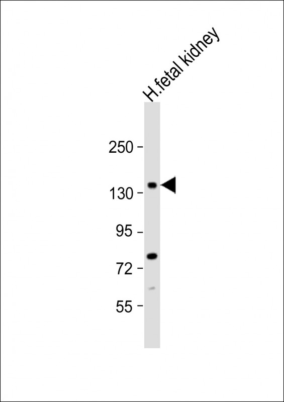
NEK1 Antibody (C-term)
Purified Rabbit Polyclonal Antibody (Pab)

- SPECIFICATION
- CITATIONS
- PROTOCOLS
- BACKGROUND

Application
| IHC-P, WB, E |
|---|---|
| Primary Accession | Q96PY6 |
| Other Accession | P51954 |
| Reactivity | Human |
| Predicted | Mouse |
| Host | Rabbit |
| Clonality | Polyclonal |
| Isotype | Rabbit IgG |
| Calculated MW | 142828 Da |
| Antigen Region | 1165-1196 aa |
| Gene ID | 4750 |
|---|---|
| Other Names | Serine/threonine-protein kinase Nek1, Never in mitosis A-related kinase 1, NimA-related protein kinase 1, Renal carcinoma antigen NY-REN-55, NEK1, KIAA1901 |
| Target/Specificity | This NEK1 antibody is generated from rabbits immunized with a KLH conjugated synthetic peptide between 1165-1196 amino acids from the C-terminal region of human NEK1. |
| Dilution | WB~~1:500 IHC-P~~1:50~100 |
| Format | Purified polyclonal antibody supplied in PBS with 0.09% (W/V) sodium azide. This antibody is prepared by Saturated Ammonium Sulfate (SAS) precipitation followed by dialysis against PBS. |
| Storage | Maintain refrigerated at 2-8°C for up to 2 weeks. For long term storage store at -20°C in small aliquots to prevent freeze-thaw cycles. |
| Precautions | NEK1 Antibody (C-term) is for research use only and not for use in diagnostic or therapeutic procedures. |
| Name | NEK1 |
|---|---|
| Synonyms | KIAA1901 |
| Function | Phosphorylates serines and threonines, but also appears to possess tyrosine kinase activity (PubMed:20230784). Involved in DNA damage checkpoint control and for proper DNA damage repair (PubMed:20230784). In response to injury that includes DNA damage, NEK1 phosphorylates VDAC1 to limit mitochondrial cell death (PubMed:20230784). May be implicated in the control of meiosis (By similarity). Involved in cilium assembly (PubMed:21211617). |
| Cellular Location | Nucleus. Cytoplasm, cytoskeleton, microtubule organizing center, centrosome {ECO:0000250|UniProtKB:P51954}. Cytoplasm. Note=Associated with the pericentriolar material (PubMed:21211617). Localizes to centrosome during interphase and mitosis (By similarity). Translocated from cytoplasm to discrete nuclear foci at sites of DNA damage (PubMed:15604234) {ECO:0000250|UniProtKB:P51954, ECO:0000269|PubMed:15604234, ECO:0000269|PubMed:21211617} |
| Tissue Location | High fetal expression in the brain and kidney. |

Thousands of laboratories across the world have published research that depended on the performance of antibodies from Abcepta to advance their research. Check out links to articles that cite our products in major peer-reviewed journals, organized by research category.
info@abcepta.com, and receive a free "I Love Antibodies" mug.

Provided below are standard protocols that you may find useful for product applications.
Background
Protein kinases are enzymes that transfer a phosphate group from a phosphate donor, generally the g phosphate of ATP, onto an acceptor amino acid in a substrate protein. By this basic mechanism, protein kinases mediate most of the signal transduction in eukaryotic cells, regulating cellular metabolism, transcription, cell cycle progression, cytoskeletal rearrangement and cell movement, apoptosis, and differentiation. With more than 500 gene products, the protein kinase family is one of the largest families of proteins in eukaryotes. The family has been classified in 8 major groups based on sequence comparison of their tyrosine (PTK) or serine/threonine (STK) kinase catalytic domains. The STE group (homologs of yeast Sterile 7, 11, 20 kinases) consists of 50 kinases related to the mitogen-activated protein kinase (MAPK) cascade families (Ste7/MAP2K, Ste11/MAP3K, and Ste20/MAP4K). MAP kinase cascades, consisting of a MAPK and one or more upstream regulatory kinases (MAPKKs) have been best characterized in the yeast pheromone response pathway. Pheromones bind to Ste cell surface receptors and activate yeast MAPK pathway.
References
Surpili, M.J., et al., Biochemistry 42(51):15369-15376 (2003).
Scanlan, M.J., et al., Int. J. Cancer 83(4):456-464 (1999).
Letwin, K., et al., EMBO J. 11(10):3521-3531 (1992).
If you have used an Abcepta product and would like to share how it has performed, please click on the "Submit Review" button and provide the requested information. Our staff will examine and post your review and contact you if needed.
If you have any additional inquiries please email technical services at tech@abcepta.com.
Ordering Information
Other Products
Shipping Information
