


 Foundational characteristics of cancer include proliferation, angiogenesis, migration, evasion of apoptosis, and cellular immortality. Find key markers for these cellular processes and antibodies to detect them.
 The SUMOplot™ Analysis Program predicts and scores sumoylation sites in your protein. SUMOylation is a post-translational modification involved in various cellular processes, such as nuclear-cytosolic transport, transcriptional regulation, apoptosis, protein stability, response to stress, and progression through the cell cycle.
 The Autophagy Receptor Motif Plotter predicts and scores autophagy receptor binding sites in your protein. Identifying proteins connected to this pathway is critical to understanding the role of autophagy in physiological as well as pathological processes such as development, differentiation, neurodegenerative diseases, stress, infection, and cancer.
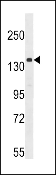
APG1 (ULK1) Antibody (Center)
Purified Rabbit Polyclonal Antibody (Pab)
_(Center)_-_U251_CQ.jpg)
- SPECIFICATION
- CITATIONS: 2
- PROTOCOLS
- BACKGROUND

Application
| WB, IF, E |
|---|---|
| Primary Accession | O75385 |
| Reactivity | Human |
| Host | Rabbit |
| Clonality | Polyclonal |
| Isotype | Rabbit IgG |
| Calculated MW | 112631 Da |
| Antigen Region | 642-672 aa |
| Gene ID | 8408 |
|---|---|
| Other Names | Serine/threonine-protein kinase ULK1, Autophagy-related protein 1 homolog, ATG1, hATG1, Unc-51-like kinase 1, ULK1, KIAA0722 |
| Target/Specificity | This APG1 (ULK1) antibody is generated from rabbits immunized with a KLH conjugated synthetic peptide between 642-672 amino acids from the Central region of human APG1 (ULK1). |
| Dilution | IF~~1:100 WB~~1:1000 |
| Format | Purified polyclonal antibody supplied in PBS with 0.09% (W/V) sodium azide. This antibody is purified through a protein A column, followed by peptide affinity purification. |
| Storage | Maintain refrigerated at 2-8°C for up to 2 weeks. For long term storage store at -20°C in small aliquots to prevent freeze-thaw cycles. |
| Precautions | APG1 (ULK1) Antibody (Center) is for research use only and not for use in diagnostic or therapeutic procedures. |
| Name | ULK1 {ECO:0000303|PubMed:9693035, ECO:0000312|HGNC:HGNC:12558} |
|---|---|
| Function | Serine/threonine-protein kinase involved in autophagy in response to starvation (PubMed:18936157, PubMed:21460634, PubMed:21795849, PubMed:23524951, PubMed:25040165, PubMed:31123703, PubMed:29487085). Acts upstream of phosphatidylinositol 3-kinase PIK3C3 to regulate the formation of autophagophores, the precursors of autophagosomes (PubMed:18936157, PubMed:21460634, PubMed:21795849, PubMed:25040165). Part of regulatory feedback loops in autophagy: acts both as a downstream effector and negative regulator of mammalian target of rapamycin complex 1 (mTORC1) via interaction with RPTOR (PubMed:21795849). Activated via phosphorylation by AMPK and also acts as a regulator of AMPK by mediating phosphorylation of AMPK subunits PRKAA1, PRKAB2 and PRKAG1, leading to negatively regulate AMPK activity (PubMed:21460634). May phosphorylate ATG13/KIAA0652 and RPTOR; however such data need additional evidences (PubMed:18936157). Plays a role early in neuronal differentiation and is required for granule cell axon formation (PubMed:11146101). Also phosphorylates SESN2 and SQSTM1 to regulate autophagy (PubMed:25040165, PubMed:37306101). Phosphorylates FLCN, promoting autophagy (PubMed:25126726). Phosphorylates AMBRA1 in response to autophagy induction, releasing AMBRA1 from the cytoskeletal docking site to induce autophagosome nucleation (PubMed:20921139). Phosphorylates ATG4B, leading to inhibit autophagy by decreasing both proteolytic activation and delipidation activities of ATG4B (PubMed:28821708). |
| Cellular Location | Cytoplasm, cytosol. Preautophagosomal structure. Note=Under starvation conditions, is localized to puncate structures primarily representing the isolation membrane that sequesters a portion of the cytoplasm resulting in the formation of an autophagosome. |
| Tissue Location | Ubiquitously expressed. Detected in the following adult tissues: skeletal muscle, heart, pancreas, brain, placenta, liver, kidney, and lung |

Provided below are standard protocols that you may find useful for product applications.
Background
Macroautophagy is the major inducible pathway for the general turnover of cytoplasmic constituents in eukaryotic cells, it is also responsible for the degradation of active cytoplasmic enzymes and organelles during nutrient starvation. Macroautophagy involves the formation of double-membrane bound autophagosomes which enclose the cytoplasmic constituent targeted for degradation in a membrane bound structure, which then fuse with the lysosome (or vacuole) releasing a single-membrane bound autophagic bodies which are then degraded within the lysosome (or vacuole). Two human homologs of the yeast autophagy-specific kinase exist: ULK1(APG1) and ULK2. APG1 plays a critical role in regulating key elements of the autophagy pathway. APG1 stimulates autophagy, leading to autophagy-dependent restriction of cell growth and ultimately cell apoptosis at high levels of activity, and is a negative regulator of mTOR signaling.
References
References for protein:
1.Scott, R., et al., Current Biology 17: 1-11 (2007).
2.Kuroyanagi, H., et al., Genomics 51(1):76-85 (1998).
References for U251 cell line:
1. Westermark B.; Pontén J.; Hugosson R. (1973).” Determinants for the establishment of permanent tissue culture lines from human gliomas”. Acta Pathol Microbiol Scand A. 81:791-805. [PMID: 4359449].
2. Pontén, J.,Westermark B. (1978).” Properties of Human Malignant Glioma Cells in Vitro”. Medical Biology 56: 184-193.[PMID: 359950].
3. Geng Y.;Kohli L.; Klocke B.J.; Roth K.A.(2010). “Chloroquine-induced autophagic vacuole accumulation and cell death in glioma cells is p53 independent”. Neuro Oncol. 12(5): 473–481.[ PMID: 20406898].
If you have used an Abcepta product and would like to share how it has performed, please click on the "Submit Review" button and provide the requested information. Our staff will examine and post your review and contact you if needed.
If you have any additional inquiries please email technical services at tech@abcepta.com.
Ordering Information
Other Products
Shipping Information
