


 Foundational characteristics of cancer include proliferation, angiogenesis, migration, evasion of apoptosis, and cellular immortality. Find key markers for these cellular processes and antibodies to detect them.
 The SUMOplot™ Analysis Program predicts and scores sumoylation sites in your protein. SUMOylation is a post-translational modification involved in various cellular processes, such as nuclear-cytosolic transport, transcriptional regulation, apoptosis, protein stability, response to stress, and progression through the cell cycle.
 The Autophagy Receptor Motif Plotter predicts and scores autophagy receptor binding sites in your protein. Identifying proteins connected to this pathway is critical to understanding the role of autophagy in physiological as well as pathological processes such as development, differentiation, neurodegenerative diseases, stress, infection, and cancer.
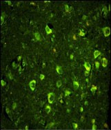
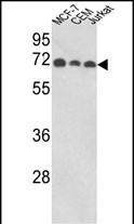
ABI1 Antibody (N-term)
Affinity Purified Rabbit Polyclonal Antibody (Pab)


- SPECIFICATION
- CITATIONS
- PROTOCOLS
- BACKGROUND

Application
| IF, FC, IHC-P, WB, E |
|---|---|
| Primary Accession | Q8IZP0 |
| Other Accession | P62484, Q9NYB9, Q9QZM5, Q8CBW3, F1R187 |
| Reactivity | Human |
| Predicted | Zebrafish, Mouse, Rat |
| Host | Rabbit |
| Clonality | Polyclonal |
| Isotype | Rabbit IgG |
| Calculated MW | 55081 Da |
| Antigen Region | 81-108 aa |
| Gene ID | 10006 |
|---|---|
| Other Names | Abl interactor 1, Abelson interactor 1, Abi-1, Abl-binding protein 4, AblBP4, Eps8 SH3 domain-binding protein, Eps8-binding protein, Nap1-binding protein, Nap1BP, Spectrin SH3 domain-binding protein 1, e3B1, ABI1, SSH3BP1 |
| Target/Specificity | This ABI1 antibody is generated from rabbits immunized with a KLH conjugated synthetic peptide between 81-108 amino acids from the N-terminal region of human ABI1. |
| Dilution | IF~~1:10~50 WB~~1:1000 IHC-P~~1:50~100 FC~~1:10~50 |
| Format | Purified polyclonal antibody supplied in PBS with 0.09% (W/V) sodium azide. This antibody is purified through a protein A column, followed by peptide affinity purification. |
| Storage | Maintain refrigerated at 2-8°C for up to 2 weeks. For long term storage store at -20°C in small aliquots to prevent freeze-thaw cycles. |
| Precautions | ABI1 Antibody (N-term) is for research use only and not for use in diagnostic or therapeutic procedures. |
| Name | ABI1 (HGNC:11320) |
|---|---|
| Synonyms | SSH3BP1 |
| Function | May act in negative regulation of cell growth and transformation by interacting with nonreceptor tyrosine kinases ABL1 and/or ABL2. May play a role in regulation of EGF-induced Erk pathway activation. Involved in cytoskeletal reorganization and EGFR signaling. Together with EPS8 participates in transduction of signals from Ras to Rac. In vitro, a trimeric complex of ABI1, EPS8 and SOS1 exhibits Rac specific guanine nucleotide exchange factor (GEF) activity and ABI1 seems to act as an adapter in the complex. Regulates ABL1/c-Abl- mediated phosphorylation of ENAH. Recruits WASF1 to lamellipodia and there seems to regulate WASF1 protein level. In brain, seems to regulate the dendritic outgrowth and branching as well as to determine the shape and number of synaptic contacts of developing neurons. |
| Cellular Location | Cytoplasm. Nucleus. Cell projection, lamellipodium. Cell projection, filopodium. Cell projection, growth cone Postsynaptic density. Cytoplasm, cytoskeleton. Note=Localized to protruding lamellipodia and filopodia tips. Also localized to neuronal growth cones and synaptosomes. May shuttle from the postsynaptic densities to the nucleus (By similarity) |
| Tissue Location | Widely expressed, with highest expression in brain. |

Thousands of laboratories across the world have published research that depended on the performance of antibodies from Abcepta to advance their research. Check out links to articles that cite our products in major peer-reviewed journals, organized by research category.
info@abcepta.com, and receive a free "I Love Antibodies" mug.

Provided below are standard protocols that you may find useful for product applications.
Background
ABI1 has been found to form a complex with EPS8 and SOS1, and is thought to be involved in the transduction of signals from Ras to Rac. In addition, this protein may play a role in the regulation of EGF-induced Erk pathway activation as well as cytoskeletal reorganization and EGFR signaling.
References
Wang,C., et.al., Mol. Cancer Res. 5 (10), 1031-1039 (2007)
Carabeo,R.A., et.al., Cell. Microbiol. 9 (9), 2278-2288 (2007)
If you have used an Abcepta product and would like to share how it has performed, please click on the "Submit Review" button and provide the requested information. Our staff will examine and post your review and contact you if needed.
If you have any additional inquiries please email technical services at tech@abcepta.com.

Ordering Information
Other Products
Shipping Information
