


 Foundational characteristics of cancer include proliferation, angiogenesis, migration, evasion of apoptosis, and cellular immortality. Find key markers for these cellular processes and antibodies to detect them.
 The SUMOplot™ Analysis Program predicts and scores sumoylation sites in your protein. SUMOylation is a post-translational modification involved in various cellular processes, such as nuclear-cytosolic transport, transcriptional regulation, apoptosis, protein stability, response to stress, and progression through the cell cycle.
 The Autophagy Receptor Motif Plotter predicts and scores autophagy receptor binding sites in your protein. Identifying proteins connected to this pathway is critical to understanding the role of autophagy in physiological as well as pathological processes such as development, differentiation, neurodegenerative diseases, stress, infection, and cancer.
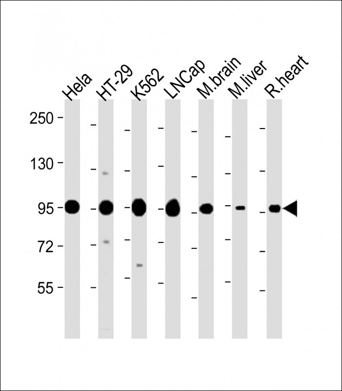
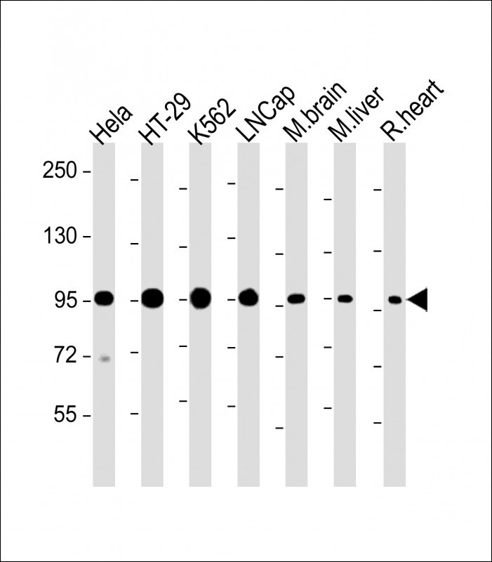
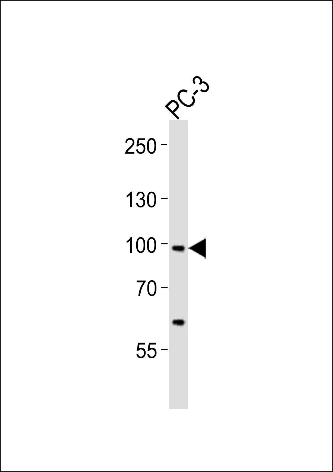
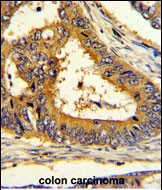
SCAP Antibody (Center)
Affinity Purified Rabbit Polyclonal Antibody (Pab)
_AP8568c_-_HeLa.jpg)
- SPECIFICATION
- CITATIONS: 1
- PROTOCOLS
- BACKGROUND

Application
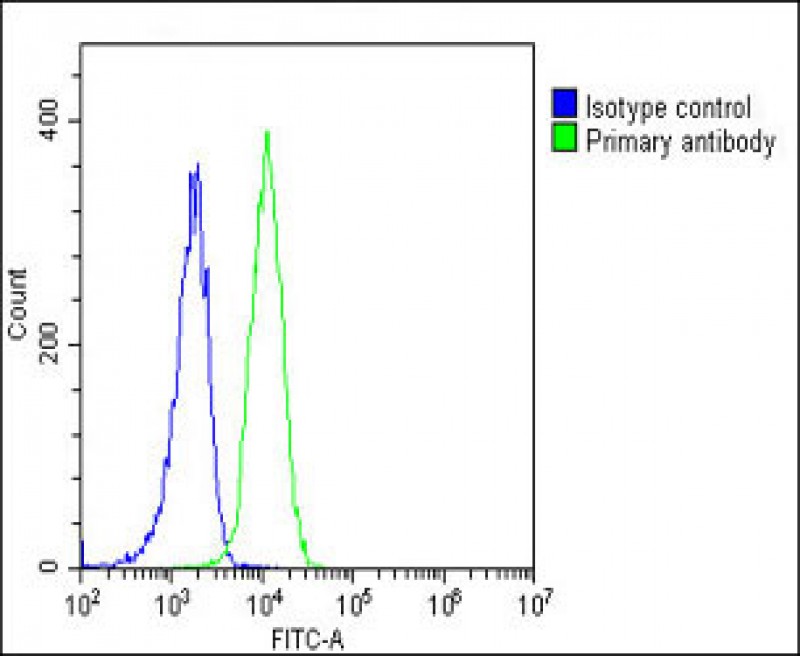
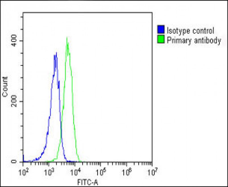
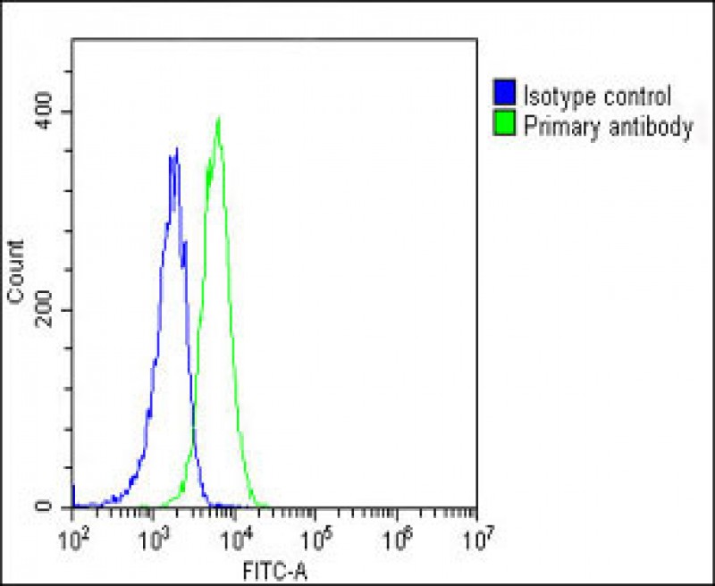
| WB, IF, IHC-P, FC, E |
|---|---|
| Primary Accession | Q12770 |
| Other Accession | A2RRU4, Q5MNU5, Q6GQT6, A6QM06 |
| Reactivity | Human, Mouse, Rat |
| Predicted | Bovine, Pig |
| Host | Rabbit |
| Clonality | Polyclonal |
| Isotype | Rabbit IgG |
| Calculated MW | 139729 Da |
| Antigen Region | 604-632 aa |
| Gene ID | 22937 |
|---|---|
| Other Names | Sterol regulatory element-binding protein cleavage-activating protein, SCAP, SREBP cleavage-activating protein, SCAP, KIAA0199 |
| Target/Specificity | This SCAP antibody is generated from rabbits immunized with a KLH conjugated synthetic peptide between 604-632 amino acids from the Central region of human SCAP. |
| Dilution | IF~~1:100 WB~~1:1000 IHC-P~~1:50~100 FC~~1:25 |
| Format | Purified polyclonal antibody supplied in PBS with 0.09% (W/V) sodium azide. This antibody is purified through a protein A column, followed by peptide affinity purification. |
| Storage | Maintain refrigerated at 2-8°C for up to 2 weeks. For long term storage store at -20°C in small aliquots to prevent freeze-thaw cycles. |
| Precautions | SCAP Antibody (Center) is for research use only and not for use in diagnostic or therapeutic procedures. |
| Name | SCAP {ECO:0000303|PubMed:10570913, ECO:0000312|HGNC:HGNC:30634} |
|---|---|
| Function | Escort protein required for cholesterol as well as lipid homeostasis (By similarity). Regulates export of the SCAP-SREBP complex from the endoplasmic reticulum to the Golgi upon low cholesterol, thereby regulating the processing of sterol regulatory element-binding proteins (SREBPs) SREBF1/SREBP1 and SREBF2/SREBP2 (By similarity). At high sterol concentrations, formation of a ternary complex with INSIG (INSIG1 or INSIG2) leads to mask the ER export signal in SCAP, promoting retention of the complex in the endoplasmic reticulum (By similarity). Low sterol concentrations trigger release of INSIG, a conformational change in the SSD domain of SCAP, unmasking of the ER export signal, promoting recruitment into COPII-coated vesicles and transport of the SCAP-SREBP to the Golgi: in the Golgi, SREBPs are then processed, releasing the transcription factor fragment of SREBPs from the membrane, its import into the nucleus and up-regulation of LDLR, INSIG1 and the mevalonate pathway (By similarity). Binds cholesterol via its SSD domain (By similarity). |
| Cellular Location | Endoplasmic reticulum membrane; Multi-pass membrane protein. Golgi apparatus membrane; Multi-pass membrane protein. Cytoplasmic vesicle, COPII- coated vesicle membrane {ECO:0000250|UniProtKB:P97260}; Multi-pass membrane protein. Note=Moves from the endoplasmic reticulum to the Golgi in the absence of sterols (By similarity) Requires the presence of SPRING1 for proper localization to endoplasmic reticulum (PubMed:32111832). {ECO:0000250|UniProtKB:P97260, ECO:0000269|PubMed:32111832} |

Provided below are standard protocols that you may find useful for product applications.
Background
SCAP is a protein with a sterol sensing domain(SSD) and seven WD domains. In the presence of cholesterol, this protein binds to sterol regulatory element binding proteins (SREBPs) and mediates their transport from the ER to the Golgi. The SREBPs are then proteolytically cleaved and regulate sterol biosynthesis.
References
Lu,Y., et.al., J. Lipid Res. 49 (12), 2582-2589 (2008)
If you have used an Abcepta product and would like to share how it has performed, please click on the "Submit Review" button and provide the requested information. Our staff will examine and post your review and contact you if needed.
If you have any additional inquiries please email technical services at tech@abcepta.com.
Ordering Information
Other Products
Shipping Information
