


 Foundational characteristics of cancer include proliferation, angiogenesis, migration, evasion of apoptosis, and cellular immortality. Find key markers for these cellular processes and antibodies to detect them.
 The SUMOplot™ Analysis Program predicts and scores sumoylation sites in your protein. SUMOylation is a post-translational modification involved in various cellular processes, such as nuclear-cytosolic transport, transcriptional regulation, apoptosis, protein stability, response to stress, and progression through the cell cycle.
 The Autophagy Receptor Motif Plotter predicts and scores autophagy receptor binding sites in your protein. Identifying proteins connected to this pathway is critical to understanding the role of autophagy in physiological as well as pathological processes such as development, differentiation, neurodegenerative diseases, stress, infection, and cancer.
INSC Antibody (Center)
Affinity Purified Rabbit Polyclonal Antibody (Pab)
- SPECIFICATION
- CITATIONS
- PROTOCOLS
- BACKGROUND

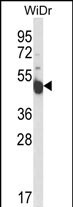
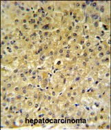
Application
| IHC-P, WB, E |
|---|---|
| Primary Accession | Q1MX18 |
| Other Accession | Q3HNM7 |
| Reactivity | Human |
| Predicted | Mouse |
| Host | Rabbit |
| Clonality | Polyclonal |
| Isotype | Rabbit IgG |
| Calculated MW | 63469 Da |
| Antigen Region | 410-437 aa |
| Gene ID | 387755 |
|---|---|
| Other Names | Protein inscuteable homolog, INSC |
| Target/Specificity | This INSC antibody is generated from rabbits immunized with a KLH conjugated synthetic peptide between 410-437 amino acids from the Central region of human INSC. |
| Dilution | WB~~1:1000 IHC-P~~1:50~100 |
| Format | Purified polyclonal antibody supplied in PBS with 0.09% (W/V) sodium azide. This antibody is purified through a protein A column, followed by peptide affinity purification. |
| Storage | Maintain refrigerated at 2-8°C for up to 2 weeks. For long term storage store at -20°C in small aliquots to prevent freeze-thaw cycles. |
| Precautions | INSC Antibody (Center) is for research use only and not for use in diagnostic or therapeutic procedures. |
| Name | INSC |
|---|---|
| Function | May function as an adapter linking the Par3 complex to the GPSM1/GPSM2 complex (PubMed:16458856). Involved in spindle orientation during mitosis. May regulate cell proliferation and differentiation in the developing nervous system. May play a role in the asymmetric division of fibroblasts and participate in the process of stratification of the squamous epithelium (By similarity). |
| Cellular Location | Cytoplasm. Cytoplasm, cell cortex. Note=Uniformly distributed in the cytoplasm during interphase. During metaphase, detected in the cell cortex, adjacent to the mitotic spindle poles |
| Tissue Location | Isoform 1 is expressed in various tissues with stronger expression in liver, kidney and small intestine. Isoform 2 is abundantly expressed in small intestine and to a lower extent in lung and pancreas. |

Thousands of laboratories across the world have published research that depended on the performance of antibodies from Abcepta to advance their research. Check out links to articles that cite our products in major peer-reviewed journals, organized by research category.
info@abcepta.com, and receive a free "I Love Antibodies" mug.

Provided below are standard protocols that you may find useful for product applications.
Background
In Drosophila, neuroblasts divide asymmetrically into another neuroblast at the apical side and a smaller ganglion mother cell on the basal side. Cell polarization is precisely regulated by 2 apically localized multiprotein signaling complexes that are tethered by Inscuteable, which regulates their apical localization (Izaki et al., 2006 [PubMed 16458856]).
References
Vural, A., et al. Mol. Cell. Biol. 30(6):1528-1540(2010)
Rivadeneira, F., et al. Nat. Genet. 41(11):1199-1206(2009)
Izaki, T., et al. Biochem. Biophys. Res. Commun. 341(4):1001-1006(2006)
Katoh, M., et al. Int. J. Mol. Med. 11(1):111-116(2003)
If you have used an Abcepta product and would like to share how it has performed, please click on the "Submit Review" button and provide the requested information. Our staff will examine and post your review and contact you if needed.
If you have any additional inquiries please email technical services at tech@abcepta.com.
Ordering Information
Other Products
Shipping Information
