


 Foundational characteristics of cancer include proliferation, angiogenesis, migration, evasion of apoptosis, and cellular immortality. Find key markers for these cellular processes and antibodies to detect them.
 The SUMOplot™ Analysis Program predicts and scores sumoylation sites in your protein. SUMOylation is a post-translational modification involved in various cellular processes, such as nuclear-cytosolic transport, transcriptional regulation, apoptosis, protein stability, response to stress, and progression through the cell cycle.
 The Autophagy Receptor Motif Plotter predicts and scores autophagy receptor binding sites in your protein. Identifying proteins connected to this pathway is critical to understanding the role of autophagy in physiological as well as pathological processes such as development, differentiation, neurodegenerative diseases, stress, infection, and cancer.
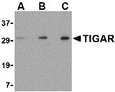
TIGAR Antibody
- SPECIFICATION
- CITATIONS
- PROTOCOLS
- BACKGROUND

Application
| WB, IHC-P, E |
|---|---|
| Primary Accession | Q9NQ88 |
| Other Accession | NP_065108, 9966849 |
| Reactivity | Human, Mouse, Rat |
| Host | Rabbit |
| Clonality | Polyclonal |
| Isotype | IgG |
| Calculated MW | 30063 Da |
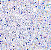
| Application Notes | TIGAR antibody can be used for detection of TIGAR by Western blot at 0.5 - 2 µg/mL. Antibody can also be used for immunohistochemistry starting at 2.5 µg/mL. |
| Gene ID | 57103 |
|---|---|
| Other Names | TIGAR Antibody: FR2BP, TIGAR, C12orf5, Fructose-2, 6-bisphosphatase TIGAR, TP53-induced glycolysis and apoptosis regulator, chromosome 12 open reading frame 5 |
| Target/Specificity | C12orf5; |
| Reconstitution & Storage | TIGAR antibody can be stored at 4℃ for three months and -20℃, stable for up to one year. As with all antibodies care should be taken to avoid repeated freeze thaw cycles. Antibodies should not be exposed to prolonged high temperatures. |
| Precautions | TIGAR Antibody is for research use only and not for use in diagnostic or therapeutic procedures. |
| Name | TIGAR {ECO:0000303|PubMed:16839880} |
|---|---|
| Synonyms | C12orf5 |
| Function | Fructose-bisphosphatase hydrolyzing fructose-2,6-bisphosphate as well as fructose-1,6-bisphosphate (PubMed:19015259). Acts as a negative regulator of glycolysis by lowering intracellular levels of fructose-2,6-bisphosphate in a p53/TP53-dependent manner, resulting in the pentose phosphate pathway (PPP) activation and NADPH production (PubMed:16839880, PubMed:22887998). Contributes to the generation of reduced glutathione to cause a decrease in intracellular reactive oxygen species (ROS) content, correlating with its ability to protect cells from oxidative or metabolic stress-induced cell death (PubMed:16839880, PubMed:19713938, PubMed:23726973, PubMed:22887998, PubMed:23817040). Plays a role in promoting protection against cell death during hypoxia by decreasing mitochondria ROS levels in a HK2- dependent manner through a mechanism that is independent of its fructose-bisphosphatase activity (PubMed:23185017). In response to cardiac damage stress, mediates p53-induced inhibition of myocyte mitophagy through ROS levels reduction and the subsequent inactivation of BNIP3. Reduced mitophagy results in an enhanced apoptotic myocyte cell death, and exacerbates cardiac damage (By similarity). Plays a role in adult intestinal regeneration; contributes to the growth, proliferation and survival of intestinal crypts following tissue ablation (PubMed:23726973). Plays a neuroprotective role against ischemic brain damage by enhancing PPP flux and preserving mitochondria functions (By similarity). Protects glioma cells from hypoxia- and ROS- induced cell death by inhibiting glycolysis and activating mitochondrial energy metabolism and oxygen consumption in a TKTL1- dependent and p53/TP53-independent manner (PubMed:22887998). Plays a role in cancer cell survival by promoting DNA repair through activating PPP flux in a CDK5-ATM-dependent signaling pathway during hypoxia and/or genome stress-induced DNA damage responses (PubMed:25928429). Involved in intestinal tumor progression (PubMed:23726973). |
| Cellular Location | Cytoplasm. Nucleus Mitochondrion. Note=Translocated to the mitochondria during hypoxia in a HIF1A-dependent manner (PubMed:23185017). Colocalizes with HK2 in the mitochondria during hypoxia (PubMed:23185017). Translocated to the nucleus during hypoxia and/or genome stress-induced DNA damage responses in cancer cells (PubMed:25928429). Translocation to the mitochondria is enhanced in ischemic cortex after reperfusion and/or during oxygen and glucose deprivation (OGD)/reoxygenation insult in primary neurons (By similarity). {ECO:0000250|UniProtKB:Q8BZA9, ECO:0000269|PubMed:23185017, ECO:0000269|PubMed:25928429} |
| Tissue Location | Expressed in the brain (PubMed:22887998). Expressed in breast tumors (PubMed:21820150). Expressed in glioblastomas (PubMed:22887998). |

Thousands of laboratories across the world have published research that depended on the performance of antibodies from Abcepta to advance their research. Check out links to articles that cite our products in major peer-reviewed journals, organized by research category.
info@abcepta.com, and receive a free "I Love Antibodies" mug.

Provided below are standard protocols that you may find useful for product applications.
Background
TIGAR Antibody: The p53 tumor-suppressor gene integrates numerous signals that control cell life and death; loss of its functions contributes to the development of most cancers. Recent studies have demonstrated the ability of p53 to regulate the expression of several proteins involved in glycolysis and oxidative phosphorylation, such as TIGAR, SCO2, and phosphoglycerate mutase. TIGAR is a recently discovered protein that functions to regulate glycolysis and protect cells against oxidative stress. TIGAR is similar in structure to proteins in the phosphoglycerate mutase family, most notably 6-phosphofructo-2-kinase, suggesting TIGAR may function as a fructose bisphosphatase. Expression of TIGAR in transfected cells correlated with an inhibition of glycolysis and decreased levels of reactive oxygen species and p53-induced apoptosis, indicating that TIGAR may act to modulate the apoptotic response to p53, thereby allowing cells to survive mild or transient stresses.
References
Guimaraes DP and Hainaut P. TP53: a key gene in human cancer. Biochimie2002; 84:83-93.
Corcoran CA, Huang Y, and Sheikh MS. The regulation of energy generating pathways by p53. Cancer Biol. Ther.2006; 5:1610-3.
Bensaad K, Tsuruta A, Selak MA, et al. TIGAR, a p53-inducible regulator of glycolysis and apoptosis. Cell2006; 126:107-20.
If you have used an Abcepta product and would like to share how it has performed, please click on the "Submit Review" button and provide the requested information. Our staff will examine and post your review and contact you if needed.
If you have any additional inquiries please email technical services at tech@abcepta.com.
Ordering Information
Other Products
Shipping Information
