


 Foundational characteristics of cancer include proliferation, angiogenesis, migration, evasion of apoptosis, and cellular immortality. Find key markers for these cellular processes and antibodies to detect them.
 The SUMOplot™ Analysis Program predicts and scores sumoylation sites in your protein. SUMOylation is a post-translational modification involved in various cellular processes, such as nuclear-cytosolic transport, transcriptional regulation, apoptosis, protein stability, response to stress, and progression through the cell cycle.
 The Autophagy Receptor Motif Plotter predicts and scores autophagy receptor binding sites in your protein. Identifying proteins connected to this pathway is critical to understanding the role of autophagy in physiological as well as pathological processes such as development, differentiation, neurodegenerative diseases, stress, infection, and cancer.
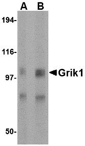
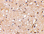
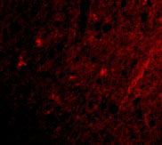
Grik1 Antibody
- SPECIFICATION
- CITATIONS
- PROTOCOLS
- BACKGROUND

Application
| WB, IHC-P, IF, E |
|---|---|
| Primary Accession | P39086 |
| Other Accession | P39086, 729597 |
| Reactivity | Human, Mouse, Rat |
| Host | Rabbit |
| Clonality | Polyclonal |
| Isotype | IgG |
| Calculated MW | 103981 Da |
| Application Notes | Grik1 antibody can be used for detection of Grik1 by Western blot at 0.5 - 1 µg/mL. Antibody can also be used for immunohistochemistry starting at 2.5 µg/mL. For immunofluorescence start at 20 µg/mL. |
| Gene ID | 2897 |
|---|---|
| Target/Specificity | GRIK1; |
| Reconstitution & Storage | Grik1 antibody can be stored at 4℃ for three months and -20℃, stable for up to one year. As with all antibodies care should be taken to avoid repeated freeze thaw cycles. Antibodies should not be exposed to prolonged high temperatures. |
| Precautions | Grik1 Antibody is for research use only and not for use in diagnostic or therapeutic procedures. |
| Name | GRIK1 |
|---|---|
| Synonyms | GLUR5 |
| Function | Ionotropic glutamate receptor. L-glutamate acts as an excitatory neurotransmitter at many synapses in the central nervous system. Binding of the excitatory neurotransmitter L-glutamate induces a conformation change, leading to the opening of the cation channel, and thereby converts the chemical signal to an electrical impulse. The receptor then desensitizes rapidly and enters a transient inactive state, characterized by the presence of bound agonist. May be involved in the transmission of light information from the retina to the hypothalamus. |
| Cellular Location | Cell membrane; Multi-pass membrane protein. Postsynaptic cell membrane; Multi-pass membrane protein |
| Tissue Location | Most abundant in the cerebellum and the suprachiasmatic nuclei (SCN) of the hypothalamus |

Thousands of laboratories across the world have published research that depended on the performance of antibodies from Abcepta to advance their research. Check out links to articles that cite our products in major peer-reviewed journals, organized by research category.
info@abcepta.com, and receive a free "I Love Antibodies" mug.

Provided below are standard protocols that you may find useful for product applications.
Background
Grik1 Antibody: Glutamate receptors are the predominant excitatory neurotransmitter receptors in the mammalian brain and are activated in a variety of normal neurophysiologic processes. Grik1, also known as glutamate receptor 5, belongs to the kainate family of glutamate receptors, which are composed of four subunits and function as ligand-activated ion channels. Grik1 is expressed in GABAergic interneurons of the hippocampus and are thought to participate in the formation of various subtypes of kainate receptors with Grik2 and KA2. Stimulation of Grik1 leads to intracellular calcium release and activation of protein kinase C. Excessive activation has been associated with psychiatric, neurological and neurodegenerative diseases. Numerous isoforms of Grik1 are known to exist and may be subject to RNA editing within the second transmembrane domain, which is thought to alter the properties of ion flow.
References
Tanaka K. Functions of glutamate transports in the brain. Neurosci. Res.2000; 37:15-9.
Pinheiro P and Mulle C. Kainate receptors. Cell Tissue Res.2006; 326:457-82.
Bureau I, Dieudonne S, Coussen F, et al. Kainate receptor-mediated responses in the CA1 field of wild-type and GluR6-deficient mice. J. Neurosci.1999; 19:653-63.
Christensen JK, Paternain AV, Selak S, et al. A mosaic of functional kainate receptors in hippocampal interneurons. J. Neurosci.2004; 24:8986-93.
If you have used an Abcepta product and would like to share how it has performed, please click on the "Submit Review" button and provide the requested information. Our staff will examine and post your review and contact you if needed.
If you have any additional inquiries please email technical services at tech@abcepta.com.
Ordering Information
Other Products
Shipping Information
