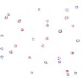
FOXO3 Antibody
- SPECIFICATION
- CITATIONS
- PROTOCOLS
- BACKGROUND

Application
| WB, IF, ICC, E |
|---|---|
| Primary Accession | O43524 |
| Other Accession | NP_963853, 42519916 |
| Reactivity | Human, Mouse, Rat |
| Host | Rabbit |
| Clonality | Polyclonal |
| Isotype | IgG |
| Calculated MW | 71277 Da |
| Application Notes | FOXO3 antibody can be used for detection of FOXO3 by Western blot at 0.5 - 1 µg/mL. Antibody can also be used for immunocytochemistry starting at 4 µg/mL. For immunofluorescence start at 20 µg/mL. |
| Gene ID | 2309 |
|---|---|
| Target/Specificity | FOXO3; |
| Reconstitution & Storage | FOXO3 antibody can be stored at 4℃ for three months and -20℃, stable for up to one year. As with all antibodies care should be taken to avoid repeated freeze thaw cycles. Antibodies should not be exposed to prolonged high temperatures. |
| Precautions | FOXO3 Antibody is for research use only and not for use in diagnostic or therapeutic procedures. |
| Name | FOXO3 (HGNC:3821) |
|---|---|
| Function | Transcriptional activator that recognizes and binds to the DNA sequence 5'-[AG]TAAA[TC]A-3' and regulates different processes, such as apoptosis and autophagy (PubMed:10102273, PubMed:16751106, PubMed:21329882, PubMed:30513302). Acts as a positive regulator of autophagy in skeletal muscle: in starved cells, enters the nucleus following dephosphorylation and binds the promoters of autophagy genes, such as GABARAP1L, MAP1LC3B and ATG12, thereby activating their expression, resulting in proteolysis of skeletal muscle proteins (By similarity). Triggers apoptosis in the absence of survival factors, including neuronal cell death upon oxidative stress (PubMed:10102273, PubMed:16751106). Participates in post-transcriptional regulation of MYC: following phosphorylation by MAPKAPK5, promotes induction of miR- 34b and miR-34c expression, 2 post-transcriptional regulators of MYC that bind to the 3'UTR of MYC transcript and prevent its translation (PubMed:21329882). In response to metabolic stress, translocates into the mitochondria where it promotes mtDNA transcription (PubMed:23283301). In response to metabolic stress, translocates into the mitochondria where it promotes mtDNA transcription. Also acts as a key regulator of chondrogenic commitment of skeletal progenitor cells in response to lipid availability: when lipids levels are low, translocates to the nucleus and promotes expression of SOX9, which induces chondrogenic commitment and suppresses fatty acid oxidation (By similarity). Also acts as a key regulator of regulatory T-cells (Treg) differentiation by activating expression of FOXP3 (PubMed:30513302). |
| Cellular Location | Cytoplasm, cytosol. Nucleus Mitochondrion matrix. Mitochondrion outer membrane; Peripheral membrane protein; Cytoplasmic side. Note=Retention in the cytoplasm contributes to its inactivation (PubMed:10102273, PubMed:15084260, PubMed:16751106). Translocates to the nucleus upon oxidative stress and in the absence of survival factors (PubMed:10102273, PubMed:16751106) Translocates from the cytosol to the nucleus following dephosphorylation in response to autophagy-inducing stimuli (By similarity). Translocates in a AMPK-dependent manner into the mitochondrion in response to metabolic stress (PubMed:23283301, PubMed:29445193). Serum deprivation increases localization to the nucleus, leading to activate expression of SOX9 and subsequent chondrogenesis (By similarity). {ECO:0000250|UniProtKB:Q9WVH4, ECO:0000269|PubMed:10102273, ECO:0000269|PubMed:15084260, ECO:0000269|PubMed:16751106, ECO:0000269|PubMed:23283301, ECO:0000269|PubMed:29445193} |
| Tissue Location | Ubiquitous.. |

Thousands of laboratories across the world have published research that depended on the performance of antibodies from Abcepta to advance their research. Check out links to articles that cite our products in major peer-reviewed journals, organized by research category.
info@abcepta.com, and receive a free "I Love Antibodies" mug.
Provided below are standard protocols that you may find useful for product applications.
Background
FOXO3 Antibody: FOXO3 is a ubiquitously expressed 75 kDa protein member of a subfamily of the forkhead homeotic gene family of transcription factors and shuttles between the cytoplasm and nucleus. FOXO transcription factors are key players of cell fate decisions, metabolism, stress resistance, tumor suppression and are regulated by growth factors, oxidative stress or nutrient deprivation. FOXO3 is involved with mTOR in the regulation of autophagy in skeletal muscle, and activates protein degradation in atrophying muscle cells. FOXO3 has also been implicated in several neurodegenerative disorders including aging, neuromuscular disease, systemic lupus erythmatosus, stroke and diabetic complications.
References
Anderson MJ, Viars CS, Czekay S, et al. Cloning and characterization of three human forkhead genes that comprise an FKHR-like gene subfamily. Genomics1998; 47:187-99.
Greer EL and Brunet A. FOXO transcription factors at the interface between longevity and tumor suppression. Oncogene2005; 24:7410-25.
Mammucari C, Schiaffino S, and Sandri M. Downstream of Akt: FoxO3 and mTOR in the regulation of autophagy in skeletal muscle. Autophagy2008; 4:524-6.
Zhao J, Brault JJ, Schild A, et al. FoxO3 coordinately activates protein degradation by the autophagic/lysosomal and proteasomal pathways in atrophying muscle cells. Cell Metab.2007; 6:472-83.
If you have used an Abcepta product and would like to share how it has performed, please click on the "Submit Review" button and provide the requested information. Our staff will examine and post your review and contact you if needed.
If you have any additional inquiries please email technical services at tech@abcepta.com.


 Foundational characteristics of cancer include proliferation, angiogenesis, migration, evasion of apoptosis, and cellular immortality. Find key markers for these cellular processes and antibodies to detect them.
 The SUMOplot™ Analysis Program predicts and scores sumoylation sites in your protein. SUMOylation is a post-translational modification involved in various cellular processes, such as nuclear-cytosolic transport, transcriptional regulation, apoptosis, protein stability, response to stress, and progression through the cell cycle.
 The Autophagy Receptor Motif Plotter predicts and scores autophagy receptor binding sites in your protein. Identifying proteins connected to this pathway is critical to understanding the role of autophagy in physiological as well as pathological processes such as development, differentiation, neurodegenerative diseases, stress, infection, and cancer.


