


 Foundational characteristics of cancer include proliferation, angiogenesis, migration, evasion of apoptosis, and cellular immortality. Find key markers for these cellular processes and antibodies to detect them.
 The SUMOplot™ Analysis Program predicts and scores sumoylation sites in your protein. SUMOylation is a post-translational modification involved in various cellular processes, such as nuclear-cytosolic transport, transcriptional regulation, apoptosis, protein stability, response to stress, and progression through the cell cycle.
 The Autophagy Receptor Motif Plotter predicts and scores autophagy receptor binding sites in your protein. Identifying proteins connected to this pathway is critical to understanding the role of autophagy in physiological as well as pathological processes such as development, differentiation, neurodegenerative diseases, stress, infection, and cancer.
DELE Antibody

- SPECIFICATION
- CITATIONS
- PROTOCOLS
- BACKGROUND

Application
| WB, IF, E |
|---|---|
| Primary Accession | Q14154 |
| Other Accession | NP_055588, 217330656 |
| Reactivity | Human, Rat |
| Host | Rabbit |
| Clonality | Polyclonal |
| Isotype | IgG |
| Calculated MW | 55920 Da |
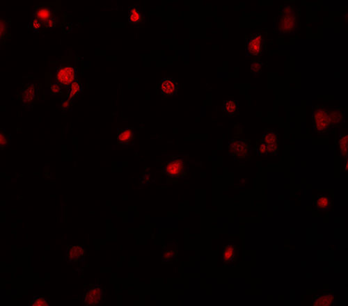
| Application Notes | DELE antibody can be used for detection of DELE by Western blot at 1 µg/mL. For immunofluorescence start at 20 µg/mL. |
| Gene ID | 9812 |
|---|---|
| Target/Specificity | KIAA0141; |
| Reconstitution & Storage | DELE antibody can be stored at 4℃ for three months and -20℃, stable for up to one year. As with all antibodies care should be taken to avoid repeated freeze thaw cycles. Antibodies should not be exposed to prolonged high temperatures. |
| Precautions | DELE Antibody is for research use only and not for use in diagnostic or therapeutic procedures. |
| Name | DELE1 (HGNC:28969) |
|---|---|
| Function | Protein kinase activator that acts as a key activator of the integrated stress response (ISR) following various stresses, such as iron deficiency and mitochondrial stress (PubMed:32132706, PubMed:32132707, PubMed:35388015, PubMed:37327776). Detects impaired protein import and processing in mitochondria, activating the ISR (PubMed:35388015). May also required for the induction of death receptor-mediated apoptosis through the regulation of caspase activation (PubMed:20563667). |
| Cellular Location | [DAP3-binding cell death enhancer 1]: Mitochondrion. Mitochondrion outer membrane. Mitochondrion inner membrane. Note=Imported in the mitochondrial matrix in absence of stress, leading to its degradation by LONP1 (PubMed:37327776). Localizes at the mitochondrial surface in response to iron deficiency: iron deficiency impairs mitochondrial import, promoting localization at the mitochondrial surface and stabilization (PubMed:37327776). Associates with the mitochondrion inner membrane in response to mitochondrial stress, leading to its proteolytic processing by OMA1, and generation of the AP3-binding cell death enhancer 1 short form (DELE1(S) or S-DELE1) (PubMed:32132707) |
| Tissue Location | Detected in liver, skeletal muscle, kidney, pancreas, spleen, thyroid, testis, ovary, small intestine and colon |

Thousands of laboratories across the world have published research that depended on the performance of antibodies from Abcepta to advance their research. Check out links to articles that cite our products in major peer-reviewed journals, organized by research category.
info@abcepta.com, and receive a free "I Love Antibodies" mug.

Provided below are standard protocols that you may find useful for product applications.
Background
DELE Antibody: DELE is a recently identified DAP3-binding protein that is thought to be important in the induction of death receptor-mediated apoptosis. Transfected cells that stably express DELE were found to be susceptible to apoptosis induction by TNF-α and TRAIL, whereas reducing DELE expression by siRNA rescued these cells from apoptosis induction. Furthermore, the reduction of DELE expression also inhibited the activation of caspase-3, caspase-8 and caspase-9 following stimulation by TNF-α, anti-Fas, or TRAIL, indicating the importance of DELE in apoptosis mediated by death receptors.
References
Harada T, Iwai A, Miyazaki T. Identification of DELE, a novel DAP-binding protein which is crucial for death receptor-mediated apoptosis induction. Apoptosis 2010; 15:1247-55.
If you have used an Abcepta product and would like to share how it has performed, please click on the "Submit Review" button and provide the requested information. Our staff will examine and post your review and contact you if needed.
If you have any additional inquiries please email technical services at tech@abcepta.com.
Ordering Information
Other Products
Shipping Information
