


 Foundational characteristics of cancer include proliferation, angiogenesis, migration, evasion of apoptosis, and cellular immortality. Find key markers for these cellular processes and antibodies to detect them.
 The SUMOplot™ Analysis Program predicts and scores sumoylation sites in your protein. SUMOylation is a post-translational modification involved in various cellular processes, such as nuclear-cytosolic transport, transcriptional regulation, apoptosis, protein stability, response to stress, and progression through the cell cycle.
 The Autophagy Receptor Motif Plotter predicts and scores autophagy receptor binding sites in your protein. Identifying proteins connected to this pathway is critical to understanding the role of autophagy in physiological as well as pathological processes such as development, differentiation, neurodegenerative diseases, stress, infection, and cancer.
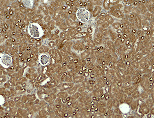
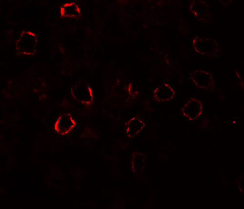
KIRREL3 Antibody

- SPECIFICATION
- CITATIONS
- PROTOCOLS
- BACKGROUND

Application
| WB, IHC-P, IF, E |
|---|---|
| Primary Accession | Q8IZU9 |
| Other Accession | NP_115920, 26006461 |
| Reactivity | Human, Mouse, Rat |
| Host | Rabbit |
| Clonality | Polyclonal |
| Isotype | IgG |
| Calculated MW | Predicted: 64, 84 kDa Observed: 60 kDa |
| Application Notes | KIRREL3 antibody can be used for detection of KIRREL3 by Western blot at 1 - 2 µg/ml. Antibody can also be used for immunohistochemistry starting at 5 µg/mL. For immunofluorescence start at 20 µg/mL. |
| Gene ID | 84623 |
|---|---|
| Target/Specificity | KIRREL3; KIRREL3 antibody is human, mouse and rat reactive. At least two isoforms are known to exist; this antibody will detect both isoforms. KIRREL3 antibody is predicted to not cross-react with other members of the KIRREL protein family. |
| Reconstitution & Storage | KIRREL3 antibody can be stored at 4℃ for three months and -20℃, stable for up to one year. |
| Precautions | KIRREL3 Antibody is for research use only and not for use in diagnostic or therapeutic procedures. |
| Name | KIRREL3 (HGNC:23204) |
|---|---|
| Function | Synaptic adhesion molecule required for the formation of target-specific synapses. Required for formation of target-specific synapses at hippocampal mossy fiber synapses. Required for formation of mossy fiber filopodia, the synaptic structures connecting dentate granule and GABA neurons. Probably acts as a homophilic adhesion molecule that promotes trans-cellular interactions and stabilize mossy fiber filipodia contact and subsequent synapse formation. Required for the coalescence of vomeronasal sensory neuron axons. May be involved in the hematopoietic supportive capacity of stroma cells; the secreted extracellular domain is directly responsible for supporting hematopoietic stem cells. |
| Cellular Location | Cell membrane; Single-pass type I membrane protein |
| Tissue Location | Expressed in fetal and adult brain (PubMed:19012874). Also expressed in kidney, specifically in podocytes of kidney glomeruli (PubMed:12424224). Also expressed in skeletal muscle (PubMed:25488023). |

Thousands of laboratories across the world have published research that depended on the performance of antibodies from Abcepta to advance their research. Check out links to articles that cite our products in major peer-reviewed journals, organized by research category.
info@abcepta.com, and receive a free "I Love Antibodies" mug.

Provided below are standard protocols that you may find useful for product applications.
Background
KIRREL3, also known as nephrin-like protein 2, is a type I transmembrane protein belonging to a family of three podocin interacting proteins and the immunoglobulin superfamily (1). KIRREL3 is involved in the regulation of both glomerular and neural development (2), and more specifically, the nucleogenesis of the pontine nuclei in the developing hindbrain (3). KIRREL3 has also been shown to interact with the synaptic scaffold protein calmodulin-associated serine/threonine kinase (CASK) in neuronal cells (4).
References
Sellin L, Huber TB, Gerke P, et al. NEPH1 defines a novel family of podocin interacting proteins. FASEB J. 2003; 17:115-7.
Neumann-Haefelin E, Kramer-Zucker A, Slanchev K, et al. A model organism approach: defining the role of Neph proteins as regulators of neuron and kidney morphogenesis. Hum. Mol. Genet. 2010; 19:2347-59.
Nishida K, Nakayama K, Yoshimura S, et al. Role of Neph2 in pontine nuclei formation in the developing hindbrain. Mol. Cell Neurosci. 2011; 46:662-70.
Mizuhara E, Minaki Y, Nakatani T, et al. Purkinje cells originate from cerebellar ventricular zone progenitors positive for Neph3 and E-cadherin. Dev. Biol. 2010; 338:202-14.
If you have used an Abcepta product and would like to share how it has performed, please click on the "Submit Review" button and provide the requested information. Our staff will examine and post your review and contact you if needed.
If you have any additional inquiries please email technical services at tech@abcepta.com.
Ordering Information
Other Products
Shipping Information
