


 Foundational characteristics of cancer include proliferation, angiogenesis, migration, evasion of apoptosis, and cellular immortality. Find key markers for these cellular processes and antibodies to detect them.
 The SUMOplot™ Analysis Program predicts and scores sumoylation sites in your protein. SUMOylation is a post-translational modification involved in various cellular processes, such as nuclear-cytosolic transport, transcriptional regulation, apoptosis, protein stability, response to stress, and progression through the cell cycle.
 The Autophagy Receptor Motif Plotter predicts and scores autophagy receptor binding sites in your protein. Identifying proteins connected to this pathway is critical to understanding the role of autophagy in physiological as well as pathological processes such as development, differentiation, neurodegenerative diseases, stress, infection, and cancer.
CaMKII (Alpha-Specific) Antibody
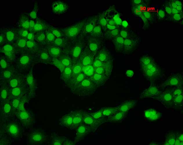
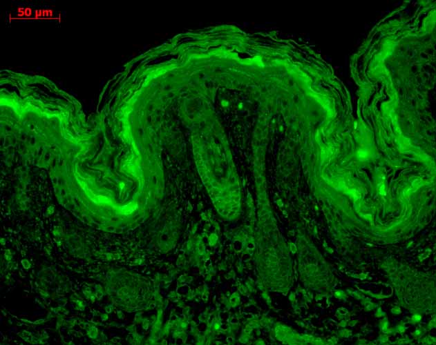
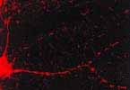
CaMKII Antibody, Clone 6G9
- SPECIFICATION
- CITATIONS
- PROTOCOLS
- BACKGROUND

Application
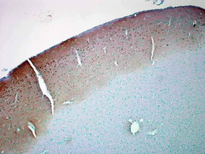
| WB, IHC, ICC/IF, IP, E, RIA |
|---|---|
| Primary Accession | P11798 |
| Other Accession | NP_033922.1 |
| Host | Mouse |
| Isotype | IgG1 |
| Reactivity | Human, Mouse, Rat, Bovine |
| Clonality | Monoclonal |
| Description | Mouse Anti-Rat CaMKII (Alpha-Specific) Monoclonal IgG1 |
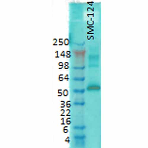
| Target/Specificity | Detects ~50-60kDa. Recognizes both phosphorylated and non-phosphorylated forms. |
| Other Names | CamK2 Antibody, CamK2A Antibody, CamK2B Antibody, CamK2D Antibody, CamK2G Antibody, CAMKA Antibody |
| Clone Names | 6G9 |
| Immunogen | Partially purified rat CaMKII |
| Purification | Protein G Purified |
| Storage | -20ºC |
| Storage Buffer | PBS pH7.4, 50% glycerol, 0.09% sodium azide |
| Shipping Temperature | Blue Ice or 4ºC |
| Certificate of Analysis | 0.1 µg/ml was sufficient for detection of CamKII in 20 µg rat brain tissue extract by colorimetric immunoblot analysis using Goat Anti-Mouse IgG:AP as the secondary. |
| Cellular Localization | Cytoplasm | Mitochondrion | Nucleus | Cell Junction | Synapse | Presynaptic Cell Membrane |

Thousands of laboratories across the world have published research that depended on the performance of antibodies from Abcepta to advance their research. Check out links to articles that cite our products in major peer-reviewed journals, organized by research category.
info@abcepta.com, and receive a free "I Love Antibodies" mug.

Provided below are standard protocols that you may find useful for product applications.
Background
CaMKII is an important member of the calcium/calmodulin-activated protein kinase family, functioning in neural synaptic stimulation and T-cell receptor signaling (1, 2). CaMKII is expressed in many different tissues but is specifically found in the neurons of the forebrain and its mRNA is found within the dendrites and the soma of the neuron. The CaMKII that is found in the neurons consist of two subunits of 52 (termed alpha genes) and 60 kDa (beta genes). CaMKII has catalytic and regulatory domains, as well as an ATP-binding domain, and a consensus phosphorylation site (3-7). The binding of Ca2+/calmodulin to its regulatory domain releases its auto inhibitory effect and activates the kinase (8). This kinase activation results in autophosphorylation at threonine 286 (8). The threonine phosphorylation state of CaMKII can be regulated through PP1/PKA. Whereas PP1 (protein phosphatase 1) dephosphorylates phospho-CaMKII at Thr286, PKA (protein kinase A) prevents this dephosphorylation (9). Autophosphorylation also enables CaMKII to attain an enhanced affinity for NMDA receptors in postsynaptic densities (10-12).
References
1. Hughes K. et al. (2001) J. Biol. Chem. 276: 36008–36013.
2. Barria A. et al. (1997) Science 276: 2042–2045.
3. Bennet M.K. and Kennedy M.B. (1987) Proc. Natl. Acad. Sci. U.S.A. 84: 1794-1798.
4. Broke L., Srinivasan M. and Schulman H. (1995) J. Neurosci. 15: 6797-6808.
5. Nghiem P., Saati S. M., Martens C. L., Gardner P. and Schulman H. (1993) J. Biol. Chem. 268: 5471-5479.
6. Edman C.F. and Schulman H. (1994) Biochem. Biophys. Acta 1221: 90-102.
7. Tombes R.M. and Krystal G.W., (1997) Biochem. Biophys. Acta 13555: 281-292.
8. Means A.R. (2000) Mol. Endocrinol. 14: 4–12.
9. Makhinson M. et al. (1999) J. Neurosci. 19: 2500–2510.
10. Strack S. and Colbran R.J. (1998) J. Biol. Chem. 273: 20689-20692.
11. Leonard S.A., Lim I.A., Hemsworth D.E., Horne M.C. and Hell J.W. (1999) Proc. Natl. Acad. Sci. U.S.A. 96: 3239-3244.
12. Shen K. and Meyer Y. (1999) Science 284: 162-167.
If you have used an Abcepta product and would like to share how it has performed, please click on the "Submit Review" button and provide the requested information. Our staff will examine and post your review and contact you if needed.
If you have any additional inquiries please email technical services at tech@abcepta.com.
Ordering Information
Other Products
Shipping Information
