


 Foundational characteristics of cancer include proliferation, angiogenesis, migration, evasion of apoptosis, and cellular immortality. Find key markers for these cellular processes and antibodies to detect them.
 The SUMOplot™ Analysis Program predicts and scores sumoylation sites in your protein. SUMOylation is a post-translational modification involved in various cellular processes, such as nuclear-cytosolic transport, transcriptional regulation, apoptosis, protein stability, response to stress, and progression through the cell cycle.
 The Autophagy Receptor Motif Plotter predicts and scores autophagy receptor binding sites in your protein. Identifying proteins connected to this pathway is critical to understanding the role of autophagy in physiological as well as pathological processes such as development, differentiation, neurodegenerative diseases, stress, infection, and cancer.
Rhodopsin Antibody
Rhodopsin Antibody, Clone 1D4
- SPECIFICATION
- CITATIONS
- PROTOCOLS
- BACKGROUND

Application
| WB, IHC, ICC/IF, IP, E |
|---|---|
| Primary Accession | P02699 |
| Other Accession | NP_001014890.1 |
| Host | Mouse |
| Isotype | IgG1 |
| Clonality | Monoclonal |
| Description | Mouse Anti-Bovine Rhodopsin Monoclonal IgG1 |
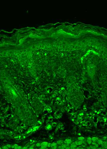
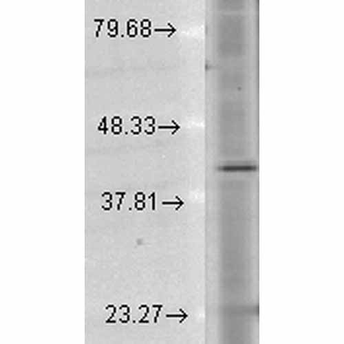
| Target/Specificity | Detects ~40kDa. Binds specifically to the N-terminus of Rhodopsin. Does not detect Rhodopsin in invertebrates. |
| Other Names | OPN2 Antibody, opsd Antibody, opsin 2 Antibody, opsin 2 rod pigment Antibody, opsin2 Antibody, RHO Antibody, RP4 Antibody, MGC138309 Antibody, Retinitis Pigmentosa 4 Antibody |
| Clone Names | 1D4 |
| Immunogen | Bovine Rhodopsin |
| Purification | Protein G Purified |
| Storage | -20ºC |
| Storage Buffer | PBS pH7.4, 50% glycerol, 0.09% sodium azide |
| Shipping Temperature | Blue Ice or 4ºC |
| Certificate of Analysis | 1 µg/ml of SMC-177 was sufficient for detection of rhodopsin in 10 µg of rat eye lysate by colorimetric immunoblot analysis using Goat anti-mouse IgG:HRP as the secondary antibody. |
| Cellular Localization | Membrane |

Thousands of laboratories across the world have published research that depended on the performance of antibodies from Abcepta to advance their research. Check out links to articles that cite our products in major peer-reviewed journals, organized by research category.
info@abcepta.com, and receive a free "I Love Antibodies" mug.

Provided below are standard protocols that you may find useful for product applications.
Background
Rhodopsin consists of the protein moiety opsin and a reversibly covalently bound cofactor, retinal. Opsin, a bundle of seven membrane embedded alpha-helices, binds retinal, a photo reactive chromophore, in a central pocket (2, 3). In addition to being the pigment of the retina that is responsible for both the formation of the photoreceptor cells, its function is to specifically convey information stored in the specific geometry of the chormophore to the surface of the molecule upon light absorption (2). In the active state, rhodopsin activates transduction, a GTP binding protein. Once activated, transduction promotes the hydrolysis of cGMP by phosphodiesterase. Rhodopsin’s activity is believed to be shut off by its phosphorylation followed by binding of the soluble protein arrestin (4). Mutations in the rhodopsin gene lead to retinitis pigmentosa, which can be inherited as an autosomal dominant, an autosomal recessive or an X-linked recessive disorder (5).
References
1. Molday R.S., Hicks D., and Molday L. (1987) Invest Ophthalmol Vis Sci. 28: 50-61.
2. Ridge K.D., Lee S.S.J., and Abdulaev N.G. (1996) J of Biol Chem. 271: 7860-7867.
3. Matsuyama T., Yamashita T., Imai H. and Shichida Y. (2009) J Biol Chem. Manuscript M109.063875.
4. Feurstein S.E., et al. (2009) Biochemistry. 48(45): 10733-10742.
5. Iannaccone A., et al. (2006) Vision Res. 46(27): 4556-4567.
If you have used an Abcepta product and would like to share how it has performed, please click on the "Submit Review" button and provide the requested information. Our staff will examine and post your review and contact you if needed.
If you have any additional inquiries please email technical services at tech@abcepta.com.
Ordering Information
Other Products
Shipping Information
