


 Foundational characteristics of cancer include proliferation, angiogenesis, migration, evasion of apoptosis, and cellular immortality. Find key markers for these cellular processes and antibodies to detect them.
 The SUMOplot™ Analysis Program predicts and scores sumoylation sites in your protein. SUMOylation is a post-translational modification involved in various cellular processes, such as nuclear-cytosolic transport, transcriptional regulation, apoptosis, protein stability, response to stress, and progression through the cell cycle.
 The Autophagy Receptor Motif Plotter predicts and scores autophagy receptor binding sites in your protein. Identifying proteins connected to this pathway is critical to understanding the role of autophagy in physiological as well as pathological processes such as development, differentiation, neurodegenerative diseases, stress, infection, and cancer.
Cav beta 2 Antibody
CavBeta2 Antibody, Clone S8B-1
- SPECIFICATION
- CITATIONS
- PROTOCOLS
- BACKGROUND

Application
| WB, IHC, ICC, IP, AM |
|---|---|
| Primary Accession | Q8VGC3 |
| Other Accession | NP_446303 |
| Host | Mouse |
| Isotype | IgG1 |
| Reactivity | Human, Mouse, Rat |
| Clonality | Monoclonal |
| Description | Mouse Anti-Rat Cav beta 2 Monoclonal IgG1 |
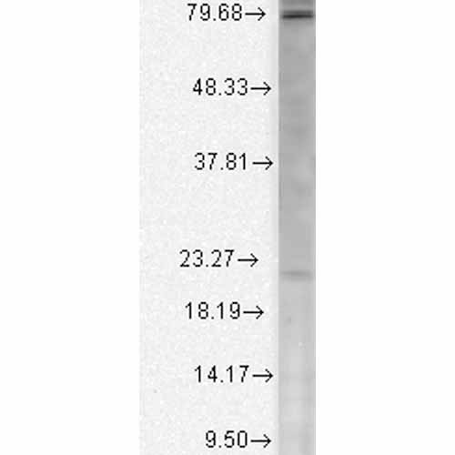
| Target/Specificity | Detects ~78 kDa. No cross reactivity against Cavβ1, Cavβ3, Cavβ4. |
| Other Names | Cacnlb2 Antibody, cacnb2 Antibody, Voltage-dependent L-type calcium channel subunit beta-2 Antibody, CAB2 Antibody |
| Clone Names | S8B-1 |
| Immunogen | Synthetic peptide amino acids 189-205 of rat CavBeta2 |
| Purification | Protein G Purified |
| Storage | -20ºC |
| Storage Buffer | PBS pH7.4, 50% glycerol, 0.09% sodium azide |
| Shipping Temperature | Blue Ice or 4ºC |
| Certificate of Analysis | 1 µg/ml of SMC-332 was sufficient for detection of Cavβ2 in 10 µg of rat brain lysate by colorimetric immunoblot analysis using Goat anti-mouse IgG:HRP as the secondary antibody |
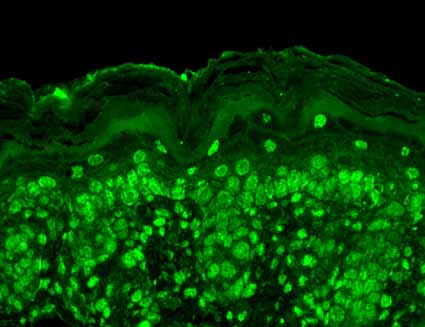
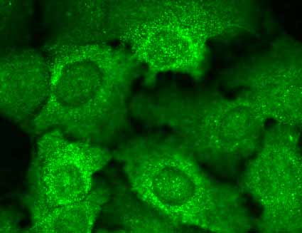
| Cellular Localization | Cell Membrane | Sarcolemma |

Thousands of laboratories across the world have published research that depended on the performance of antibodies from Abcepta to advance their research. Check out links to articles that cite our products in major peer-reviewed journals, organized by research category.
info@abcepta.com, and receive a free "I Love Antibodies" mug.

Provided below are standard protocols that you may find useful for product applications.
Background
Cav Beat subunits are involved in the transport of the pore forming alpha1 subunit to the plasma membrane (1). They also shield an ER Retention signal on the alpha1 subunit, thereby guiding the pore-forming subunit to the target membrane (2, 3). They also determine the biophysical properties of the calcium channel (3).
References
1. Dolphin A.C. (2003) J Bioenerg. Biomembr. 35: 599-620.
2. Bichet D., et al. (2000) Neuron. 25: 177-190.
3. Xie M., et al. (2007) J Cell Biol. 178(3): 489-502.
If you have used an Abcepta product and would like to share how it has performed, please click on the "Submit Review" button and provide the requested information. Our staff will examine and post your review and contact you if needed.
If you have any additional inquiries please email technical services at tech@abcepta.com.
Ordering Information
Shipping Information
