


 Foundational characteristics of cancer include proliferation, angiogenesis, migration, evasion of apoptosis, and cellular immortality. Find key markers for these cellular processes and antibodies to detect them.
 The SUMOplot™ Analysis Program predicts and scores sumoylation sites in your protein. SUMOylation is a post-translational modification involved in various cellular processes, such as nuclear-cytosolic transport, transcriptional regulation, apoptosis, protein stability, response to stress, and progression through the cell cycle.
 The Autophagy Receptor Motif Plotter predicts and scores autophagy receptor binding sites in your protein. Identifying proteins connected to this pathway is critical to understanding the role of autophagy in physiological as well as pathological processes such as development, differentiation, neurodegenerative diseases, stress, infection, and cancer.
Ankyrin R Antibody
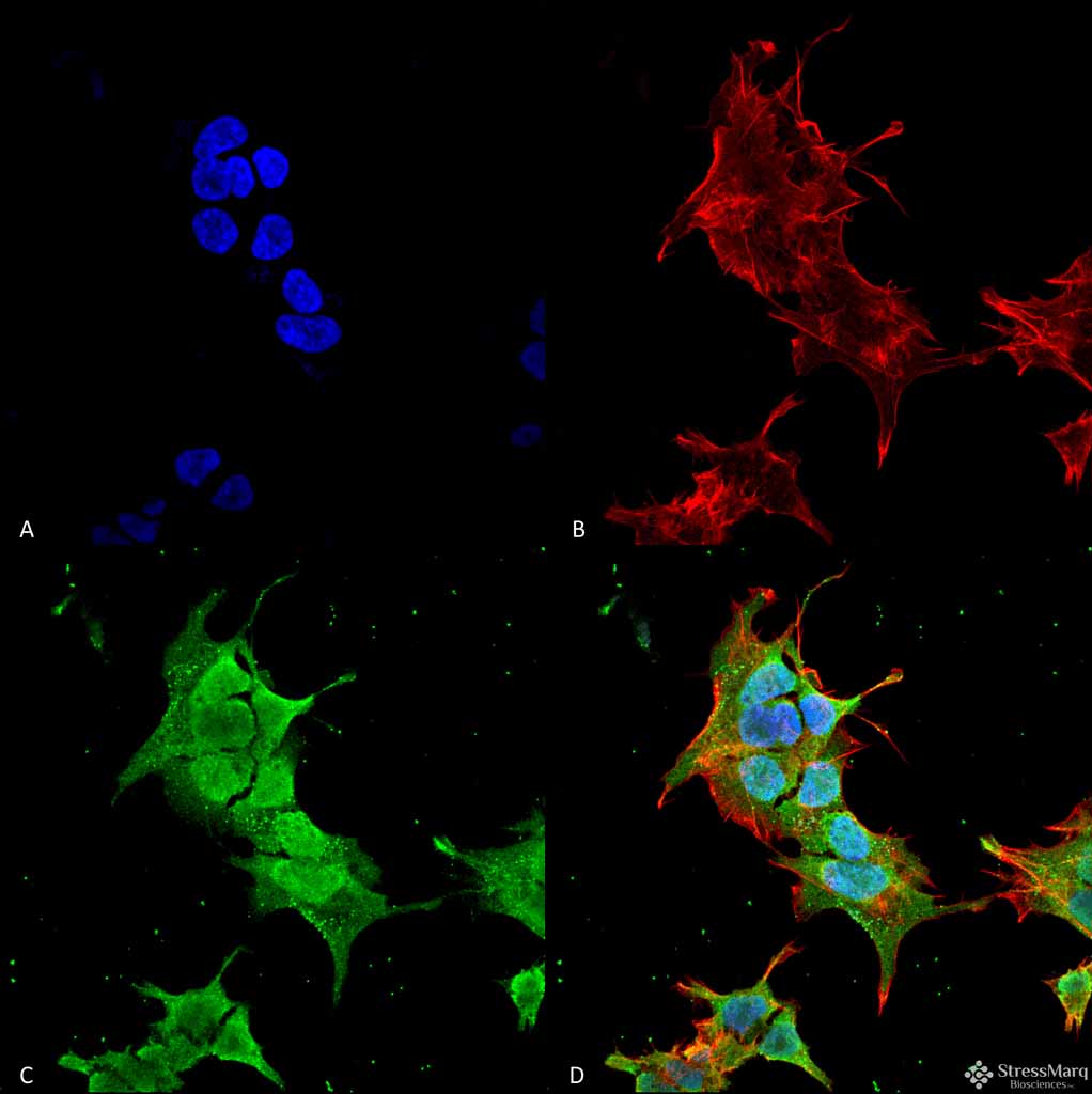
Ankyrin R Antibody, Clone S388A-10
- SPECIFICATION
- CITATIONS
- PROTOCOLS
- BACKGROUND

Application
| WB, IHC, ICC |
|---|---|
| Primary Accession | P16157 |
| Other Accession | NP_000028.3 |
| Host | Mouse |
| Isotype | IgG2B |
| Reactivity | Human, Mouse, Rat |
| Clonality | Monoclonal |
| Description | Mouse Anti-Human Ankyrin R Monoclonal IgG2B |
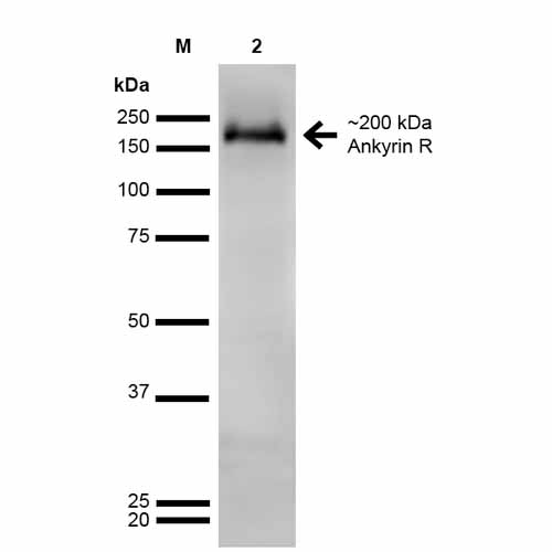
| Target/Specificity | Detects ~200kDa. Does not cross-react with Ankyrin-B or Ankyrin-G. |
| Other Names | Ankyrin-R Antibody, Ankyrin-1 Antibody, Erythrocyte Ankyrin Antibody, ANK-1 Antibody, ANK Antibody, rCG_43073 Antibody |
| Clone Names | S388A-10 |
| Immunogen | Fusion protein amino acids 1-1881 (full-length) of human Ankyrin-R |
| Purification | Protein G Purified |
| Storage | -20ºC |
| Storage Buffer | PBS pH7.4, 50% glycerol, 0.1% sodium azide |
| Shipping Temperature | Blue Ice or 4ºC |
| Certificate of Analysis | A 1:100 dilution of SMC-487 was sufficient for detection of Ankyrin R in 20 µg of mouse brain lysate by ECL immunoblot analysis using Goat anti-mouse IgG:HRP as the secondary antibody. |
| Cellular Localization | Cytoplasm | Cytoskeleton | Myofibril | Sarcomere | M line |

Thousands of laboratories across the world have published research that depended on the performance of antibodies from Abcepta to advance their research. Check out links to articles that cite our products in major peer-reviewed journals, organized by research category.
info@abcepta.com, and receive a free "I Love Antibodies" mug.

Provided below are standard protocols that you may find useful for product applications.
Background
Ankyrins are a family of adaptor proteins that mediate the attachment of integral membrane proteins to the spectrin-actin based membrane skeleton (1). Ankyrins have binding sites for the beta subunit of spectrin and at least 12 families of integral membrane proteins. This linkage is required to maintain the integrity of the plasma membranes and to anchor specific ion channels, ion exchangers and ion transporters in the plasma membrane. Ankyrin R, or Ank1, was first discovered in erythrocyres, but has since also been located in the brain and muscles. Mutations are associated with hereditary spehrocytosis and alzheimer's diease (2).
References
1. Bennett V., Baines A.J. (2001) Physiol. Rev. 81 (3): 1353–92.
2. Genes and mapped phenotypes. (n.d.). Retrieved April 21, 2015, from http://www.ncbi.nlm.nih.gov/gene/286
If you have used an Abcepta product and would like to share how it has performed, please click on the "Submit Review" button and provide the requested information. Our staff will examine and post your review and contact you if needed.
If you have any additional inquiries please email technical services at tech@abcepta.com.
Ordering Information
Other Products
Shipping Information
