


 Foundational characteristics of cancer include proliferation, angiogenesis, migration, evasion of apoptosis, and cellular immortality. Find key markers for these cellular processes and antibodies to detect them.
 The SUMOplot™ Analysis Program predicts and scores sumoylation sites in your protein. SUMOylation is a post-translational modification involved in various cellular processes, such as nuclear-cytosolic transport, transcriptional regulation, apoptosis, protein stability, response to stress, and progression through the cell cycle.
 The Autophagy Receptor Motif Plotter predicts and scores autophagy receptor binding sites in your protein. Identifying proteins connected to this pathway is critical to understanding the role of autophagy in physiological as well as pathological processes such as development, differentiation, neurodegenerative diseases, stress, infection, and cancer.
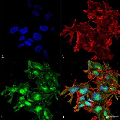
ATG2A Antibody: ATTO 390
- SPECIFICATION
- CITATIONS
- PROTOCOLS
- BACKGROUND

Application
| WB, ICC/IF |
|---|---|
| Primary Accession | Q2TAZ0 |
| Other Accession | NP_055919.2 |
| Host | Rabbit |
| Reactivity | Human |
| Clonality | Polyclonal |
| Description | Rabbit Anti-Human ATG2A Polyclonal |
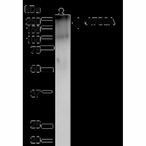
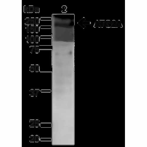
| Target/Specificity | Predicted molecular weights at ~213kDa. Other observed bands around ~90-100kDa to be expected. |
| Other Names | ATG2 autophagy related homolog A Antibody, ATG2a_HUMAN Antibody, BC023754 Antibody, KIAA0404 Antibody, 1810013C15Rik Antibody |
| Immunogen | Synthetic peptide from the C-terminal of human ATG2A |
| Purification | Peptide Affinity Purified |
| Storage | -20ºC |
| Storage Buffer | PBS, 50% glycerol, 0.09% sodium azide |
| Shipping Temperature | Blue Ice or 4ºC |
| Certificate of Analysis | A 1:1000 dilution of SPC-652 was sufficient for detection of ATG2A on HeLa cell lysates using Goat anti-rabbit IgG:HRP as the secondary antibody. |
| Cellular Localization | Preautophagosomal Structure Membrane | Peripheral membrane protein |

Thousands of laboratories across the world have published research that depended on the performance of antibodies from Abcepta to advance their research. Check out links to articles that cite our products in major peer-reviewed journals, organized by research category.
info@abcepta.com, and receive a free "I Love Antibodies" mug.

Provided below are standard protocols that you may find useful for product applications.
Background
ATG2A belongs to the ATG2 family. It is required for both autophagosome formation and regulation of lipid droplet morphology and dispersion (1).
References
1. Velikkakath A.K., Nishimura T., Oita E., Ishihara N., Mizushima N. (2012) Mol Biol Cell. 23(5): 896-909.
If you have used an Abcepta product and would like to share how it has performed, please click on the "Submit Review" button and provide the requested information. Our staff will examine and post your review and contact you if needed.
If you have any additional inquiries please email technical services at tech@abcepta.com.
Ordering Information
Other Products
Shipping Information
