


 Foundational characteristics of cancer include proliferation, angiogenesis, migration, evasion of apoptosis, and cellular immortality. Find key markers for these cellular processes and antibodies to detect them.
 The SUMOplot™ Analysis Program predicts and scores sumoylation sites in your protein. SUMOylation is a post-translational modification involved in various cellular processes, such as nuclear-cytosolic transport, transcriptional regulation, apoptosis, protein stability, response to stress, and progression through the cell cycle.
 The Autophagy Receptor Motif Plotter predicts and scores autophagy receptor binding sites in your protein. Identifying proteins connected to this pathway is critical to understanding the role of autophagy in physiological as well as pathological processes such as development, differentiation, neurodegenerative diseases, stress, infection, and cancer.
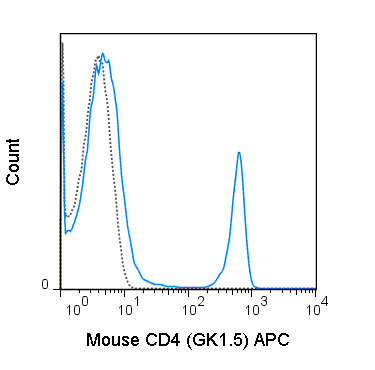
APC Anti-Mouse CD4 (GK1.5) Antibody
- SPECIFICATION
- CITATIONS
- PROTOCOLS
- BACKGROUND

Application
| FC |
|---|---|
| Isotype | Rat IgG2b, kappa |
| Concentration | 0.2 mg/mL |
| Reactivity | Mouse |
| Formulation | 10 mM NaH2PO4, 150 mM NaCl, 0.09% NaN3, 0.1% gelatin, pH7.2 |
| Host | Rat |
| Gene ID | 12504 |
|---|---|
| Gene Name | Cd4 |
| Alternative Name(s) | L3T4, T4 |
| Format | APC |
| Preparation | This monoclonal antibody was purified from tissue culture supernatant via affinity chromatography. The purified antibody was conjugated under optimal conditions, with unreacted dye removed from the preparation. It is recommended to store the product undiluted at 4°C, and protected from prolonged exposure to light. Do not freeze. |
| Application Notes | This antibody preparation has been quality-tested for flow cytometry using mouse spleen cells, or an appropriate cell type (where indicated). The amount of antibody required for optimal staining of a cell sample should be determined empirically in your system. |
| Storage Conditions | 2-8°C protected from light |
Taniguchi RT, DeVoss JJ, Moon JJ, Sidney J, Sette A, Jenkins MK, and Anderson MS. 2012. Proc. Natl. Acad. Sci. 109: 7847-7852. (in vivo depletion)
Wolkers MC, Gerlach, C, Arens R, Janssen EM, Fitzgerald P, Schumacher TN, Medema JP, Green DR, and Schoenberger SP. 2012. Blood. 119:798-804. (in vivo depletion)
Lee L-F, Logronio K, Tu GH, Zhai W, Ni I, Mei L, Dilley J, Yu J, et al. 2012. Proc. Natl. Acad. Sci. 10.1073. (Flow cytometry).
Thornton EE, Looney MR, Bose O, Sen D, Sheppard D, Locksley R, Huang X, and Krummel MF. 2012. J. Exp. Med. 10:1084. (Immunohistochemistry – OCT embedded frozen tissue)
Stephen TL, Wilson BS, and Laufer TM. 2012. Proc. Natl. Acad. Sci. 109: 7415-7420. (Immunoprecipitation)
Hammerbeck CD and Hooper JW. 2011. J. Virol. 85(19) 9929-9944. (Flow cytometry – Syrian hamster).
Diamond MS, Kinder M, Matsushita H, Mashayekhi M, Dunn GP, Archambault JM, Lee H, Arthur CD, White JM, Kalinke U, Murphy KM, and Schreiber RD. 2011. J. Exp. Med. 208: 1989-2003. (in vitro blocking)
Kao H, Lin J, Littman DR, Shaw AS, and Allen PM. 2008. J. Immunol. 181: 8248-8257. (Immunoprecipitation)
Felix NJ, Donermeyer DL, Horvath S, Walters JJ, Gross M, suri A, and Allen PM. 2007. Nat. Immunol. 8:388-397. (in vitro blocking)
Provided below are standard protocols that you may find useful for product applications.
Background
The GK1.5 antibody reacts with mouse CD4, a 55 kDa protein which acts as a co-receptor for the T cell receptor (TCR) in its interaction with MHC Class II molecules on antigen-presenting cells. The extracellular domain of CD4 binds to the beta-2 domain of MHC Class II, while its cytoplasmic tail provides a binding site for the tyrosine kinase lck, facilitating the signaling cascade that initiates T cell activation. CD4 is typically expressed on thymocytes, certain mature T cell populations such as Th17 and T regulatory (Treg) cells, as well as on dendritic cells.
The GK1.5 antibody is widely used as a phenotypic marker for CD4 expression. If used together, the GK1.5 antibody and an alternative antibody, Anti-Mouse CD4 clone RM4-5, will “compete” for binding, i.e. RM4-5 is able to block GK1.5 binding to cells. In contrast, the Anti-Mouse CD4 clone RM4-4 does not block binding of the GK1.5 antibody to cells (Arora S et al. 2006. Infect. Immun. 74: 4339-4348). The GK1.5 antibody is also reported to be cross-reactive with Syrian hamster CD4.

If you have used an Abcepta product and would like to share how it has performed, please click on the "Submit Review" button and provide the requested information. Our staff will examine and post your review and contact you if needed.
If you have any additional inquiries please email technical services at tech@abcepta.com.
Ordering Information
Other Products
Shipping Information
