


 Foundational characteristics of cancer include proliferation, angiogenesis, migration, evasion of apoptosis, and cellular immortality. Find key markers for these cellular processes and antibodies to detect them.
 The SUMOplot™ Analysis Program predicts and scores sumoylation sites in your protein. SUMOylation is a post-translational modification involved in various cellular processes, such as nuclear-cytosolic transport, transcriptional regulation, apoptosis, protein stability, response to stress, and progression through the cell cycle.
 The Autophagy Receptor Motif Plotter predicts and scores autophagy receptor binding sites in your protein. Identifying proteins connected to this pathway is critical to understanding the role of autophagy in physiological as well as pathological processes such as development, differentiation, neurodegenerative diseases, stress, infection, and cancer.
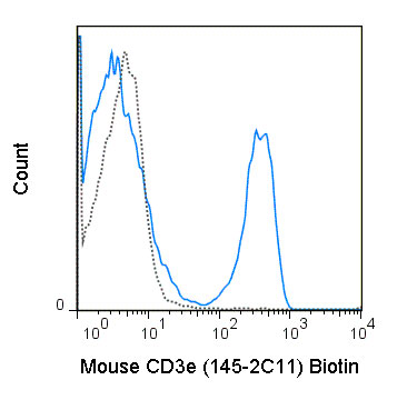
Biotin Anti-Mouse CD3e (145-2C11) Antibody
- SPECIFICATION
- CITATIONS
- PROTOCOLS
- BACKGROUND

Application
| FC |
|---|---|
| Isotype | Armenian Hamster IgG |
| Concentration | 0.5 mg/mL |
| Reactivity | Mouse |
| Formulation | 10 mM NaH2PO4, 150 mM NaCl, 0.09% NaN3, 0.1% gelatin, pH7.2 |
| Host | Armenian Hamster |
| Gene ID | 12501 |
|---|---|
| Gene Name | Cd3e |
| Alternative Name(s) | CD3 epsilon |
| Format | Biotin |
| Preparation | This monoclonal antibody was purified from tissue culture supernatant via affinity chromatography. The purified antibody was conjugated under optimal conditions, with unreacted dye removed from the preparation. It is recommended to store the product undiluted at 4°C, and protected from prolonged exposure to light. Do not freeze. |
| Application Notes | This antibody preparation has been quality-tested for flow cytometry using mouse spleen cells, or an appropriate cell type (where indicated). The amount of antibody required for optimal staining of a cell sample should be determined empirically in your system. |
| Storage Conditions | 2-8°C protected from light |
Staehli R, Ludigs K, Heinz LX, Sequin-Estevez Q, Ferero I, Braun M, Schroder K, Rebsamen M, Tardivel A, Mattmann C, MacDonald HR, Romero P, Reith W, Guarda G, and Tschopp J. 2012. J. Immunol. 188: 3820-3828. (in vitro activation)
Todo T, Wu G, Chai NN, He Y, Martins G, Gupta A, Fair J, Liu NY, Jordan S, and Klein A. 2012. Int. Immunol. 10:1093. (in vivo assay)
Mira E, Leon B, Barber DF, Jimenez-Baranda S, Goya I, Almonacid L, Marquez G, Zaballos A, Martinez C, Stein JV, Ardavin C and Manes S. 2012. J. Immunol. (in vitro activation, Immunohistochemistry – frozen tissue)
Becker-Herman A, Meyer-Bahlburg A, Schwartz MA, Jackson SW, Hudkins KL, Liu C, Sather BD, Khim S, Liggitt D, Song W, Silverman GJ, Alpers CE and Rawlings DJ. 2011. J. Exp. Med. 208:2033-2042. (Immunofluorescence microscopy – OCT embedded frozen tissue)
Salmond RJ, Filby A, Pirinen N, Magee AI, and Zamoyska R. 2010. Blood. 117: 108-117. (Immunoprecipitation)
Tilley SL, Jaradat M, Stapleton C, Dixon D, Hua X, Erikson CJ,. McCaskill JG, Chason KD, Liao G, Jania L, Koller BH, and Jetten AM. 2007. J. Immunol. 178: 3208-18. (Immunohistochemistry – frozen tissue)
Isakov N, Wange RL, Burgess WH, Watts JD, Aebersold R, and Samelson LE. 1995. J. Exp. Med. 181:375-380. (in vitro activation, Immunoprecipitation)
Salvadori S, Gansbacher B, Pizzimenti AM, and Zier KS. 1994. J. Immunol. 153: 5176 - 5182. (Western Blotting)
Leo O, Foo M, Sachs D, Samuelson L, and Bluestone J. 1987. Proc. Natl. Acad. Sci. USA. 84: 1374 (Origination of clone 145-2C11, in vitro activation, in vitro blocking, Immunoprecipitation)
Provided below are standard protocols that you may find useful for product applications.
Background
The 145-2C11 antibody is specific for mouse CD3e, also known as CD3 epsilon, a 20 kDa subunit of the T cell receptor complex, along with CD3 gamma and CD3 delta. These integral membrane protein chains assemble with additional chains of the T cell receptor (TCR), as well as CD3 zeta chain, to form the T cell receptor – CD3 complex. Together with co-receptors CD4 or CD8, the complex serves to recognize antigens bound to MHC molecules on antigen-presenting cells. Such interactions promote T cell receptor signaling (T cell activation) and can result in a number of cellular responses including proliferation, differentiation, production of cytokines or activation-induced cell death. CD3 is differentially expressed during thymocyte-to-T cell development and on all mature T cells.
The 145-2C11 antibody is a widely used phenotypic marker for mouse T cells. In addition, binding of 145-2C11 antibody to CD3e can induce cell activation. A recent publication of the crystal structure of a murine CD3e-mitogenic antibody complex provides further insight into the action of commonly used agonist antibodies (Fernandes, R.A. et al. 2012. J. Biol. Chem. 287: 13324-13335).

If you have used an Abcepta product and would like to share how it has performed, please click on the "Submit Review" button and provide the requested information. Our staff will examine and post your review and contact you if needed.
If you have any additional inquiries please email technical services at tech@abcepta.com.
Ordering Information
Other Products
Shipping Information
