


 Foundational characteristics of cancer include proliferation, angiogenesis, migration, evasion of apoptosis, and cellular immortality. Find key markers for these cellular processes and antibodies to detect them.
 The SUMOplot™ Analysis Program predicts and scores sumoylation sites in your protein. SUMOylation is a post-translational modification involved in various cellular processes, such as nuclear-cytosolic transport, transcriptional regulation, apoptosis, protein stability, response to stress, and progression through the cell cycle.
 The Autophagy Receptor Motif Plotter predicts and scores autophagy receptor binding sites in your protein. Identifying proteins connected to this pathway is critical to understanding the role of autophagy in physiological as well as pathological processes such as development, differentiation, neurodegenerative diseases, stress, infection, and cancer.
Anti-RANK Picoband Antibody
- SPECIFICATION
- CITATIONS
- PROTOCOLS
- BACKGROUND

Application
| WB, IHC-P |
|---|---|
| Primary Accession | Q9Y6Q6 |
| Host | Rabbit |
| Reactivity | Human, Mouse, Rat |
| Clonality | Polyclonal |
| Format | Lyophilized |
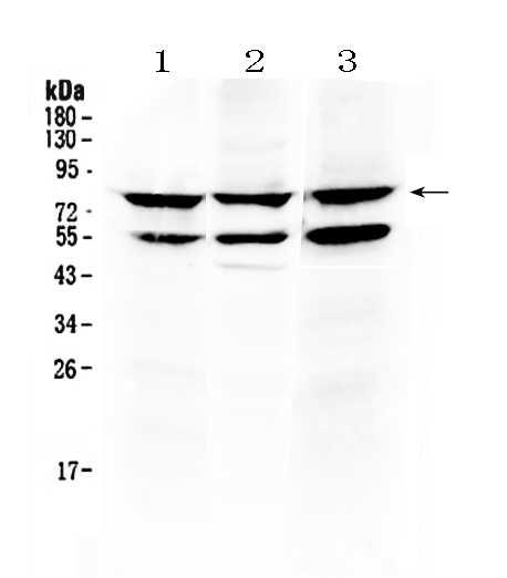
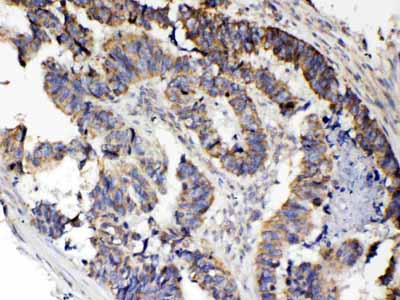
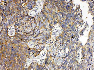
| Description | Rabbit IgG polyclonal antibody for Tumor necrosis factor receptor superfamily member 11A(TNFRSF11A) detection. Tested with WB, IHC-P in Human;Mouse;Rat. |
| Reconstitution | Add 0.2ml of distilled water will yield a concentration of 500ug/ml. |
| Gene ID | 8792 |
|---|---|
| Other Names | Tumor necrosis factor receptor superfamily member 11A, Osteoclast differentiation factor receptor, ODFR, Receptor activator of NF-KB, CD265, TNFRSF11A, RANK |
| Calculated MW | 66034 MW KDa |
| Application Details | Immunohistochemistry(Paraffin-embedded Section), 0.5-1 µg/ml, Human, By Heat Western blot, 0.1-0.5 µg/ml, Human, Mouse, Rat |
| Subcellular Localization | Isoform 1: Cell membrane ; Single-pass type I membrane protein . |
| Tissue Specificity | Ubiquitous expression with high levels in skeletal muscle, thymus, liver, colon, small intestine and adrenal gland. |
| Protein Name | Tumor necrosis factor receptor superfamily member 11A |
| Contents | Each vial contains 5mg BSA, 0.9mg NaCl, 0.2mg Na2HPO4, 0.05mg NaN3. |
| Immunogen | A synthetic peptide corresponding to a sequence in the middle region of human RANK (235-262aa YRKKGKALTANLWHWINEACGRLSGDKE), different from the related mouse sequence by seven amino acids. |
| Purification | Immunogen affinity purified. |
| Cross Reactivity | No cross reactivity with other proteins. |
| Storage | At -20˚C for one year. After r˚Constitution, at 4˚C for one month. It˚Can also be aliquotted and stored frozen at -20˚C for a longer time.Avoid repeated freezing and thawing. |
| Name | TNFRSF11A |
|---|---|
| Synonyms | RANK |
| Function | Receptor for TNFSF11/RANKL/TRANCE/OPGL; essential for RANKL- mediated osteoclastogenesis (PubMed:9878548). Its interaction with EEIG1 promotes osteoclastogenesis via facilitating the transcription of NFATC1 and activation of PLCG2 (By similarity). Involved in the regulation of interactions between T-cells and dendritic cells (By similarity). |
| Cellular Location | [Isoform 1]: Cell membrane; Single-pass type I membrane protein. Membrane raft {ECO:0000250|UniProtKB:O35305} |
| Tissue Location | Ubiquitous expression with high levels in skeletal muscle, thymus, liver, colon, small intestine and adrenal gland |

Thousands of laboratories across the world have published research that depended on the performance of antibodies from Abcepta to advance their research. Check out links to articles that cite our products in major peer-reviewed journals, organized by research category.
info@abcepta.com, and receive a free "I Love Antibodies" mug.

Provided below are standard protocols that you may find useful for product applications.
Background
Receptor Activator of Nuclear Factor κ B (RANK), also known as TRANCE Receptor, is a type I membrane protein that is expressed on the surface of osteoclasts and is involved in their activation upon ligand binding. RANK is also expressed on dendritic cells and facilitates immune signaling. It is found on the surface of stromal cells, osteoblasts, and T cells. By analysis of somatic cell and radiation hybrid panels, this gene is mapped to 18q22.1.
If you have used an Abcepta product and would like to share how it has performed, please click on the "Submit Review" button and provide the requested information. Our staff will examine and post your review and contact you if needed.
If you have any additional inquiries please email technical services at tech@abcepta.com.
Ordering Information
Other Products
Shipping Information
