


 Foundational characteristics of cancer include proliferation, angiogenesis, migration, evasion of apoptosis, and cellular immortality. Find key markers for these cellular processes and antibodies to detect them.
 The SUMOplot™ Analysis Program predicts and scores sumoylation sites in your protein. SUMOylation is a post-translational modification involved in various cellular processes, such as nuclear-cytosolic transport, transcriptional regulation, apoptosis, protein stability, response to stress, and progression through the cell cycle.
 The Autophagy Receptor Motif Plotter predicts and scores autophagy receptor binding sites in your protein. Identifying proteins connected to this pathway is critical to understanding the role of autophagy in physiological as well as pathological processes such as development, differentiation, neurodegenerative diseases, stress, infection, and cancer.
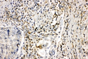
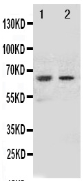
Anti-NOX1 Antibody
- SPECIFICATION
- CITATIONS
- PROTOCOLS
- BACKGROUND

Application
| WB, IHC-P |
|---|---|
| Primary Accession | Q9Y5S8 |
| Host | Rabbit |
| Reactivity | Human |
| Clonality | Polyclonal |
| Format | Lyophilized |
| Description | Rabbit IgG polyclonal antibody for NADPH oxidase 1(NOX1) detection. Tested with WB, IHC-P in Human. |
| Reconstitution | Add 0.2ml of distilled water will yield a concentration of 500ug/ml. |
| Gene ID | 27035 |
|---|---|
| Other Names | NADPH oxidase 1, NOX-1, 1.-.-.-, Mitogenic oxidase 1, MOX-1, NADH/NADPH mitogenic oxidase subunit P65-MOX, NOH-1, NOX1, MOX1, NOH1 |
| Calculated MW | 64871 MW KDa |
| Application Details | Immunohistochemistry(Paraffin-embedded Section), 0.5-1 µg/ml, Human, By Heat Western blot, 0.1-0.5 µg/ml, Human |
| Subcellular Localization | Cell projection, invadopodium membrane ; Multi-pass membrane protein . |
| Tissue Specificity | NOH-1L is detected in colon, uterus, prostate, and colon carcinoma, but not in peripheral blood leukocytes. NOH- 1S is detected only in colon and colon carcinoma cells. |
| Protein Name | NADPH oxidase 1(NOX-1) |
| Contents | Each vial contains 5mg BSA, 0.9mg NaCl, 0.2mg Na2HPO4, 0.05mg Thimerosal, 0.05mg NaN3. |
| Immunogen | A synthetic peptide corresponding to a sequence in the middle region of human NOX1(354-374aa, HIRAAGDWTENLIRAFEQQYS). |
| Purification | Immunogen affinity purified. |
| Cross Reactivity | No cross reactivity with other proteins |
| Storage | At -20˚C for one year. After r˚Constitution, at 4˚C for one month. It˚Can also be aliquotted and stored frozen at -20˚C for a longer time.Avoid repeated freezing and thawing. |
| Sequence Similarities | Contains 1 FAD-binding FR-type domain. |
| Name | NOX1 |
|---|---|
| Synonyms | MOX1, NOH1 |
| Function | NADPH oxidase that catalyzes the generation of superoxide from molecular oxygen utilizing NADPH as an electron donor. |
| Cellular Location | Cell projection, invadopodium membrane; Multi-pass membrane protein. Cell membrane; Multi-pass membrane protein |
| Tissue Location | [Isoform NOH-1L]: Detected in colon, uterus, prostate, and colon carcinoma, but not in peripheral blood leukocytes |

Thousands of laboratories across the world have published research that depended on the performance of antibodies from Abcepta to advance their research. Check out links to articles that cite our products in major peer-reviewed journals, organized by research category.
info@abcepta.com, and receive a free "I Love Antibodies" mug.

Provided below are standard protocols that you may find useful for product applications.
Background
NOX1(NADPH OXIDASE 1), also known as NOH1, MOX1 or GP91-2, is an enzyme that in humans is encoded by the NOX1 gene. It is also a homolog of the catalytic subunit of the superoxide-generating NADPH oxidase of phagocytes, gp91phox. The NOX1 gene is mapped to Xq22.1. NOX1 was expressed in colon, prostate, uterus, and vascular smooth muscle, but not in peripheral blood leukocytes. The deduced 564-amino acid NOX1 protein, which is 58% identical to CYBB, contains 6 membrane-spanning regions, conserved flavin and pyridine nucleotide-binding sites, and histidines possibly involved in heme ligation. Overexpression of MOX1 in NIH 3T3 cells increased superoxide generation and cell growth. Cells expressing MOX1 had a transformed appearance, showed anchorage-independent growth, and produced tumors in athymic mice. Disruption of either Nox1 or Nox2 significantly delayed progression of motor neuron disease in these mice. However, 50% survival rates were enhanced significantly more by Nox2 deletion than Nox1 deletion.
If you have used an Abcepta product and would like to share how it has performed, please click on the "Submit Review" button and provide the requested information. Our staff will examine and post your review and contact you if needed.
If you have any additional inquiries please email technical services at tech@abcepta.com.
Ordering Information
Other Products
Shipping Information
