


 Foundational characteristics of cancer include proliferation, angiogenesis, migration, evasion of apoptosis, and cellular immortality. Find key markers for these cellular processes and antibodies to detect them.
 The SUMOplot™ Analysis Program predicts and scores sumoylation sites in your protein. SUMOylation is a post-translational modification involved in various cellular processes, such as nuclear-cytosolic transport, transcriptional regulation, apoptosis, protein stability, response to stress, and progression through the cell cycle.
 The Autophagy Receptor Motif Plotter predicts and scores autophagy receptor binding sites in your protein. Identifying proteins connected to this pathway is critical to understanding the role of autophagy in physiological as well as pathological processes such as development, differentiation, neurodegenerative diseases, stress, infection, and cancer.
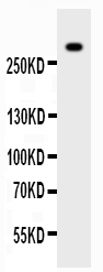
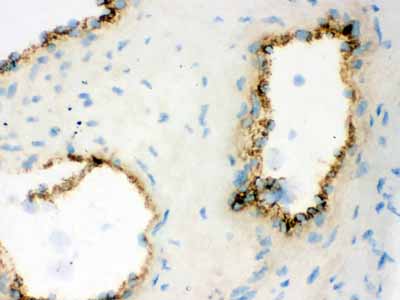
Anti-VWF Picoband Antibody

- SPECIFICATION
- CITATIONS
- PROTOCOLS
- BACKGROUND

Application
| WB, IHC-P, IHC-F |
|---|---|
| Primary Accession | P04275 |
| Host | Rabbit |
| Reactivity | Human, Mouse |
| Clonality | Polyclonal |
| Format | Lyophilized |
| Description | Rabbit IgG polyclonal antibody for von Willebrand factor(VWF) detection. Tested with WB, IHC-P, IHC-F in Human;Mouse. |
| Reconstitution | Add 0.2ml of distilled water will yield a concentration of 500ug/ml. |
| Gene ID | 7450 |
|---|---|
| Other Names | von Willebrand factor, vWF, von Willebrand antigen 2, von Willebrand antigen II, VWF, F8VWF |
| Calculated MW | 309265 MW KDa |
| Application Details | Immunohistochemistry(Paraffin-embedded Section), 0.5-1 µg/ml, By Heat Immunohistochemistry(Frozen Section), 0.5-1 µg/ml Western blot, 0.1-0.5 µg/ml |
| Subcellular Localization | Secreted . Secreted, extracellular space, extracellular matrix . Localized to storage granules. |
| Tissue Specificity | Plasma. |
| Protein Name | von Willebrand factor |
| Contents | Each vial contains 5mg BSA, 0.9mg NaCl, 0.2mg Na2HPO4, 0.05mg NaN3. |
| Immunogen | E.coli-derived human VWF recombinant protein (Position: R2535-K2813). Human VWF shares 79% amino acid (aa) sequence identity with mouse VWF. |
| Purification | Immunogen affinity purified. |
| Cross Reactivity | No cross reactivity with other proteins |
| Storage | At -20˚C for one year. After r˚Constitution, at 4˚C for one month. It˚Can also be aliquotted and stored frozen at -20˚C for a longer time.Avoid repeated freezing and thawing. |
| Sequence Similarities | Contains 1 CTCK (C-terminal cystine knot-like) domain. |
| Name | VWF |
|---|---|
| Synonyms | F8VWF |
| Function | Important in the maintenance of hemostasis, it promotes adhesion of platelets to the sites of vascular injury by forming a molecular bridge between sub-endothelial collagen matrix and platelet- surface receptor complex GPIb-IX-V. Also acts as a chaperone for coagulation factor VIII, delivering it to the site of injury, stabilizing its heterodimeric structure and protecting it from premature clearance from plasma. |
| Cellular Location | Secreted. Secreted, extracellular space, extracellular matrix. Note=Localized to storage granules |
| Tissue Location | Plasma. |

Thousands of laboratories across the world have published research that depended on the performance of antibodies from Abcepta to advance their research. Check out links to articles that cite our products in major peer-reviewed journals, organized by research category.
info@abcepta.com, and receive a free "I Love Antibodies" mug.

Provided below are standard protocols that you may find useful for product applications.
Background
Von Willebrand factor (VWF) is a blood glycoprotein involved in hemostasis. It is mapped to 12p13.31. The VWF gene encodes von Willebrand factor (VWF), a large multimeric glycoprotein that plays a central role in the blood coagulation system, serving both as a major mediator of platelet-vessel wall interaction and platelet adhesion, and as a carrier for coagulation factor VIII. VWF released from endothelial cell Weibel-Palade bodies bound particularly avidly to the extracellular matrix. VWF deficiency or dysfunction (von Willebrand disease) leads to a bleeding tendency, which is most apparent in tissues having high blood flow shear in narrow vessels.
If you have used an Abcepta product and would like to share how it has performed, please click on the "Submit Review" button and provide the requested information. Our staff will examine and post your review and contact you if needed.
If you have any additional inquiries please email technical services at tech@abcepta.com.
Ordering Information
Other Products
Shipping Information
