


 Foundational characteristics of cancer include proliferation, angiogenesis, migration, evasion of apoptosis, and cellular immortality. Find key markers for these cellular processes and antibodies to detect them.
 The SUMOplot™ Analysis Program predicts and scores sumoylation sites in your protein. SUMOylation is a post-translational modification involved in various cellular processes, such as nuclear-cytosolic transport, transcriptional regulation, apoptosis, protein stability, response to stress, and progression through the cell cycle.
 The Autophagy Receptor Motif Plotter predicts and scores autophagy receptor binding sites in your protein. Identifying proteins connected to this pathway is critical to understanding the role of autophagy in physiological as well as pathological processes such as development, differentiation, neurodegenerative diseases, stress, infection, and cancer.
Anti-SLC2A1 Picoband Antibody


- SPECIFICATION
- CITATIONS
- PROTOCOLS
- BACKGROUND

Application
| WB, IHC-P, IHC-F, ICC |
|---|---|
| Primary Accession | P11166 |
| Host | Rabbit |
| Reactivity | Human, Mouse, Rat |
| Clonality | Polyclonal |
| Format | Lyophilized |
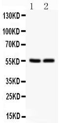
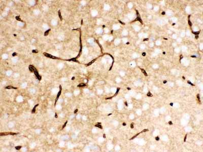
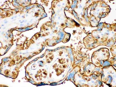
| Description | Rabbit IgG polyclonal antibody for Solute carrier family 2, facilitated glucose transporter member 1(SLC2A1) detection. Tested with WB, IHC-P, IHC-F, ICC in Human;Mouse;Rat. |
| Reconstitution | Add 0.2ml of distilled water will yield a concentration of 500ug/ml. |
| Gene ID | 6513 |
|---|---|
| Other Names | Solute carrier family 2, facilitated glucose transporter member 1, Glucose transporter type 1, erythrocyte/brain, GLUT-1, HepG2 glucose transporter, SLC2A1, GLUT1 |
| Calculated MW | 54084 MW KDa |
| Application Details | Immunocytochemistry , 0.5-1 µg/ml, Human, - Immunohistochemistry(Frozen Section), 0.5-1 µg/ml, Human, - Immunohistochemistry(Paraffin-embedded Section), 0.5-1 µg/ml, Human, Mouse, Rat, By Heat Western blot, 0.1-0.5 µg/ml, Human |
| Subcellular Localization | Cell membrane; Multi-pass membrane protein. Melanosome. Localizes primarily at the cell surface. Identified by mass spectrometry in melanosome fractions from stage I to stage IV. |
| Tissue Specificity | Detected in erythrocytes (at protein level). Expressed at variable levels in many human tissues. . |
| Protein Name | Solute carrier family 2, facilitated glucose transporter member 1 |
| Contents | Each vial contains 5mg BSA, 0.9mg NaCl, 0.2mg Na2HPO4, 0.05mg NaN3. |
| Immunogen | E.coli-derived human SLC2A1 recombinant protein (Position: R92-V492). Human SLC2A1 shares 98% and 98.3% amino acid (aa) sequence identity with mouse and rat SLC2A1, respectively. |
| Purification | Immunogen affinity purified. |
| Cross Reactivity | No cross reactivity with other proteins |
| Storage | At -20˚C for one year. After r˚Constitution, at 4˚C for one month. It˚Can also be aliquotted and stored frozen at -20˚C for a longer time.Avoid repeated freezing and thawing. |
| Sequence Similarities | Belongs to the major facilitator superfamily. Sugar transporter (TC 2.A.1.1) family. Glucose transporter subfamily. |
| Name | SLC2A1 (HGNC:11005) |
|---|---|
| Function | Facilitative glucose transporter, which is responsible for constitutive or basal glucose uptake (PubMed:18245775, PubMed:19449892, PubMed:25982116, PubMed:27078104, PubMed:10227690). Has a very broad substrate specificity; can transport a wide range of aldoses including both pentoses and hexoses (PubMed:18245775, PubMed:19449892). Most important energy carrier of the brain: present at the blood-brain barrier and assures the energy-independent, facilitative transport of glucose into the brain (PubMed:10227690). In association with BSG and NXNL1, promotes retinal cone survival by increasing glucose uptake into photoreceptors (By similarity). Required for mesendoderm differentiation (By similarity). |
| Cellular Location | Cell membrane; Multi-pass membrane protein. Melanosome. Photoreceptor inner segment {ECO:0000250|UniProtKB:P17809}. Note=Localizes primarily at the cell surface (PubMed:18245775, PubMed:19449892, PubMed:23219802, PubMed:25982116, PubMed:24847886). Identified by mass spectrometry in melanosome fractions from stage I to stage IV (PubMed:17081065) |
| Tissue Location | Detected in erythrocytes (at protein level). Expressed at variable levels in many human tissues |

Thousands of laboratories across the world have published research that depended on the performance of antibodies from Abcepta to advance their research. Check out links to articles that cite our products in major peer-reviewed journals, organized by research category.
info@abcepta.com, and receive a free "I Love Antibodies" mug.

Provided below are standard protocols that you may find useful for product applications.
Background
GLUT1, also known as SLC2A1, is a major glucose transporter in the mammalian blood-brain barrier whose gene is mapped to 1p35-p31.3 and contains 10 exons. It is present at high levels in primate erythrocytes and brain endothelial cells. Not only can transport dehydroascorbic acid (the oxidized form of vitamin C) into the brain, GLUT1 is also likely to contribute to HTLV-associated disorders through interacting with HTLV envelope glycoproteins. Functionally, GLUT1 deficiency causes a decrease in embryonic glucose uptake and apoptosis, which may be involved in diabetic embryopathy, by contrast, an increased expression of GLUT1 in some malignant tumors may suggest a role for glucose-derivative tracers to detect in vivo thyroid cancer metastases by positron-emission tomography scanning.
If you have used an Abcepta product and would like to share how it has performed, please click on the "Submit Review" button and provide the requested information. Our staff will examine and post your review and contact you if needed.
If you have any additional inquiries please email technical services at tech@abcepta.com.
Ordering Information
Other Products
Shipping Information
