


 Foundational characteristics of cancer include proliferation, angiogenesis, migration, evasion of apoptosis, and cellular immortality. Find key markers for these cellular processes and antibodies to detect them.
 The SUMOplot™ Analysis Program predicts and scores sumoylation sites in your protein. SUMOylation is a post-translational modification involved in various cellular processes, such as nuclear-cytosolic transport, transcriptional regulation, apoptosis, protein stability, response to stress, and progression through the cell cycle.
 The Autophagy Receptor Motif Plotter predicts and scores autophagy receptor binding sites in your protein. Identifying proteins connected to this pathway is critical to understanding the role of autophagy in physiological as well as pathological processes such as development, differentiation, neurodegenerative diseases, stress, infection, and cancer.
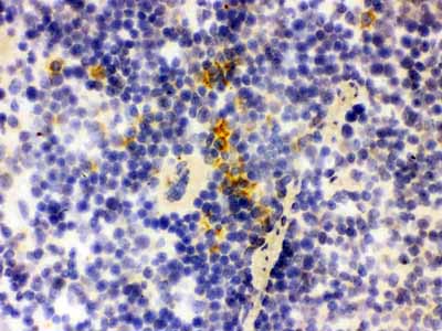
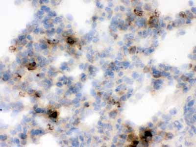
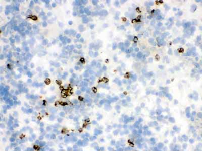
Anti-Lipocalin-2/NGAL Antibody
- SPECIFICATION
- CITATIONS
- PROTOCOLS
- BACKGROUND

Application
| WB, IHC-P, IHC-F, E |
|---|---|
| Primary Accession | P30152 |
| Host | Rabbit |
| Reactivity | Mouse, Rat |
| Clonality | Polyclonal |
| Format | Lyophilized |
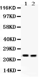
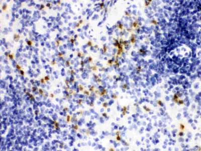
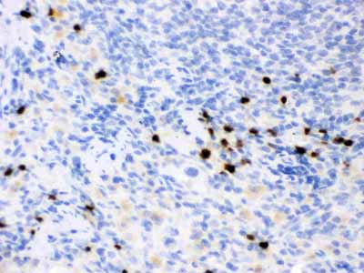
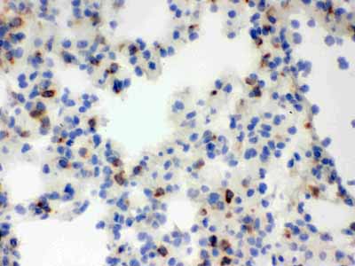
| Description | Rabbit IgG polyclonal antibody for Neutrophil gelatinase-associated lipocalin(LCN2) detection. Tested with WB, IHC-P, IHC-F, ELISA in Mouse; Rat. |
| Reconstitution | Add 0.2ml of distilled water will yield a concentration of 500ug/ml. |
| Gene ID | 170496 |
|---|---|
| Other Names | Neutrophil gelatinase-associated lipocalin, NGAL, Alpha-2-microglobulin-related protein, Alpha-2U globulin-related protein, Lipocalin-2, Siderocalin LCN2, p25, Lcn2 |
| Calculated MW | 22476 MW KDa |
| Application Details | Immunohistochemistry(Paraffin-embedded Section), 0.5-1 µg/ml, By Heat Immunohistochemistry(Frozen Section), 0.5-1 µg/ml ELISA , 0.1-0.5 µg/ml Western blot, 0.1-0.5 µg/ml |
| Subcellular Localization | Secreted . Upon binding to the SLC22A17 (24p3R) receptor, it is internalized. . |
| Protein Name | Neutrophil gelatinase-associated lipocalin |
| Contents | Each vial contains 5mg BSA, 0.9mg NaCl, 0.2mg Na2HPO4, 0.05mg NaN3. |
| Immunogen | E. coli-derived rat Lipocalin 2 recombinant protein (Position: Q21-N198). Rat Lipocalin 2 shares 64.4 % and 81.1 % amino acid (aa) sequence identity with human and mouse Lipocalin 2, respectively. |
| Purification | Immunogen affinity purified. |
| Cross Reactivity | No cross reactivity with other proteins |
| Storage | At -20˚C for one year. After r˚Constitution, at 4˚C for one month. It˚Can also be aliquotted and stored frozen at -20˚C for a longer time.Avoid repeated freezing and thawing. |
| Name | Lcn2 |
|---|---|
| Function | Iron-trafficking protein involved in multiple processes such as apoptosis, innate immunity and renal development (By similarity). Binds iron through association with 2,3-dihydroxybenzoic acid (2,3- DHBA), a siderophore that shares structural similarities with bacterial enterobactin, and delivers or removes iron from the cell, depending on the context. Iron-bound form (holo-24p3) is internalized following binding to the SLC22A17 (24p3R) receptor, leading to release of iron and subsequent increase of intracellular iron concentration. In contrast, association of the iron-free form (apo-24p3) with the SLC22A17 (24p3R) receptor is followed by association with an intracellular siderophore, iron chelation and iron transfer to the extracellular medium, thereby reducing intracellular iron concentration. Involved in apoptosis due to interleukin-3 (IL3) deprivation: iron-loaded form increases intracellular iron concentration without promoting apoptosis, while iron-free form decreases intracellular iron levels, inducing expression of the proapoptotic protein BCL2L11/BIM, resulting in apoptosis (By similarity). Involved in innate immunity; limits bacterial proliferation by sequestering iron bound to microbial siderophores, such as enterobactin. Can also bind siderophores from M.tuberculosis (By similarity). |
| Cellular Location | Secreted {ECO:0000250|UniProtKB:P80188}. Cytoplasmic granule lumen {ECO:0000250|UniProtKB:P80188}. Cytoplasmic vesicle lumen {ECO:0000250|UniProtKB:P80188}. Note=Upon binding to the SLC22A17 (24p3R) receptor, it is internalized (By similarity). Releases the bound iron in the acidic lumen of cytoplasmic vesicles (By similarity). {ECO:0000250|UniProtKB:P11672, ECO:0000250|UniProtKB:P80188} |
| Tissue Location | Detected in the ureteric bud in embryonic kidney (at protein level). |

Thousands of laboratories across the world have published research that depended on the performance of antibodies from Abcepta to advance their research. Check out links to articles that cite our products in major peer-reviewed journals, organized by research category.
info@abcepta.com, and receive a free "I Love Antibodies" mug.

Provided below are standard protocols that you may find useful for product applications.
Background
Europhile gelatinase-associated lipocalin (NGAL) is a protein that in humans is encoded by the LCN2 gene. The binding of lipocalin-2 to bacterial siderophores is important in the innate immune response to bacterial infection. Upon encountering invading bacteria the toll-like receptors on immune cells stimulate the synthesis and secretion of lipocalin-2. Secreted lipocalin-2 then limits bacterial growth by sequestering iron-containing siderophores. Lipocalin-2 also functions as a growth factor.
If you have used an Abcepta product and would like to share how it has performed, please click on the "Submit Review" button and provide the requested information. Our staff will examine and post your review and contact you if needed.
If you have any additional inquiries please email technical services at tech@abcepta.com.
Ordering Information
Shipping Information
