


 Foundational characteristics of cancer include proliferation, angiogenesis, migration, evasion of apoptosis, and cellular immortality. Find key markers for these cellular processes and antibodies to detect them.
 The SUMOplot™ Analysis Program predicts and scores sumoylation sites in your protein. SUMOylation is a post-translational modification involved in various cellular processes, such as nuclear-cytosolic transport, transcriptional regulation, apoptosis, protein stability, response to stress, and progression through the cell cycle.
 The Autophagy Receptor Motif Plotter predicts and scores autophagy receptor binding sites in your protein. Identifying proteins connected to this pathway is critical to understanding the role of autophagy in physiological as well as pathological processes such as development, differentiation, neurodegenerative diseases, stress, infection, and cancer.
ILK1 Antibody
Rabbit Polyclonal Antibody
- SPECIFICATION
- CITATIONS
- PROTOCOLS
- BACKGROUND

Application
| WB, IHC |
|---|---|
| Primary Accession | O55222 |
| Other Accession | NP_034692 |
| Reactivity | Human, Mouse, Rat |
| Host | Rabbit |
| Clonality | Polyclonal |
| Isotype | Rabbit IgG |
| Calculated MW | 51373 Da |
| Gene ID | 16202 |
|---|---|
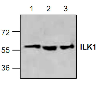
| Application & Usage | Western blotting (0.5-4 µg/ml), Immunohistochemistry (10-20 µg/ml, frozen & paraffin). However, the optimal concentrations should be determined individually. The antibody recognizes 59 kDa of ILK1 from samples of human, mouse, and rat origins. Reactivity to other species has not been determined. |
| Other Names | p59ILK , ILK-1 , P59 , DKFZp686F1765 |
| Target/Specificity | ILK1 |
| Antibody Form | Liquid |
| Appearance | Colorless liquid |
| Formulation | 100 µg (0.5 mg/ml) affinity purified rabbit polyclonal antibody in phosphate-buffered saline (PBS) containing 30% glycerol, 0.5% BSA, and 0.01% thimerosal. |
| Handling | The antibody solution should be gently mixed before use. |
| Reconstitution & Storage | -20 °C |
| Background Descriptions | |
| Precautions | ILK1 Antibody is for research use only and not for use in diagnostic or therapeutic procedures. |
| Name | Ilk {ECO:0000312|MGI:MGI:1195267} |
|---|---|
| Function | Receptor-proximal protein kinase regulating integrin-mediated signal transduction. May act as a mediator of inside-out integrin signaling. Focal adhesion protein part of the complex ILK-PINCH. This complex is considered to be one of the convergence points of integrin- and growth factor-signaling pathway. Could be implicated in mediating cell architecture, adhesion to integrin substrates and anchorage-dependent growth in epithelial cells. Regulates cell motility by forming a complex with PARVB. Phosphorylates beta-1 and beta-3 integrin subunit on serine and threonine residues, but also AKT1 and GSK3B. |
| Cellular Location | Cell junction, focal adhesion. Cell membrane; Peripheral membrane protein; Cytoplasmic side {ECO:0000250|UniProtKB:Q13418}. Cytoplasm, myofibril, sarcomere {ECO:0000250|UniProtKB:Q13418}. Cell projection, lamellipodium |
| Tissue Location | Highly expressed in lung, heart, kidney, liver, brain, spleen and skeletal muscle. Weakly expressed in testis |

Thousands of laboratories across the world have published research that depended on the performance of antibodies from Abcepta to advance their research. Check out links to articles that cite our products in major peer-reviewed journals, organized by research category.
info@abcepta.com, and receive a free "I Love Antibodies" mug.

Provided below are standard protocols that you may find useful for product applications.
Background
ILKs (Integrin-linked kinases) in combination with integrins and growth factors regulate cell survival, cell cycle, cell-cell adhesion and cell motility. ILK functions as a scaffold bridging the extra-cellular matrix (ECM) and growth factor receptors to the actin cytoskeleton thro µgh interactions with integrin, PINCH (which links ILK to the RTKs via Nck2), CH-ILKBP and affixin. ILK phosphorylates several cellular targets including Akt, GSK-3, myosin light chain 2, as well as affixin. These phosphorylation events are key regulatory steps in modulating the activities of the targets.
If you have used an Abcepta product and would like to share how it has performed, please click on the "Submit Review" button and provide the requested information. Our staff will examine and post your review and contact you if needed.
If you have any additional inquiries please email technical services at tech@abcepta.com.
Ordering Information
Other Products
Shipping Information
