


 Foundational characteristics of cancer include proliferation, angiogenesis, migration, evasion of apoptosis, and cellular immortality. Find key markers for these cellular processes and antibodies to detect them.
 The SUMOplot™ Analysis Program predicts and scores sumoylation sites in your protein. SUMOylation is a post-translational modification involved in various cellular processes, such as nuclear-cytosolic transport, transcriptional regulation, apoptosis, protein stability, response to stress, and progression through the cell cycle.
 The Autophagy Receptor Motif Plotter predicts and scores autophagy receptor binding sites in your protein. Identifying proteins connected to this pathway is critical to understanding the role of autophagy in physiological as well as pathological processes such as development, differentiation, neurodegenerative diseases, stress, infection, and cancer.
HDAC10 Antibody
Rabbit Polyclonal Antibody
- SPECIFICATION
- CITATIONS
- PROTOCOLS
- BACKGROUND

Application
| WB |
|---|---|
| Primary Accession | Q969S8 |
| Other Accession | AAL30513 |
| Reactivity | Human, Mouse, Rat |
| Host | Rabbit |
| Clonality | Polyclonal |
| Isotype | Rabbit IgG |
| Calculated MW | 71445 Da |
| Gene ID | 83933 |
|---|---|
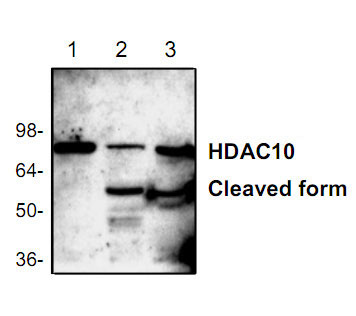
| Application & Usage | Western blotting (0.5-4 µg/ml), However, the optimal concentrations should be determined individually. The antibody recognizes 74 kDa HDAC-10 of human, mouse, and rat origins. A 55 kDa cleavage fragment can also be detected in mouse and rat tissue lysates. |
| Other Names | HD10 , Histone deacetylase 10 |
| Target/Specificity | HDAC10 |
| Antibody Form | Liquid |
| Appearance | Colorless liquid |
| Formulation | 100 µg (0.2 mg/ml) affinity purified rabbit polyclonal antibody in phosphate-buffered saline (PBS) containing 30% glycerol, 0.5% BSA, and 0.01% thimerosal. |
| Handling | The antibody solution should be gently mixed before use. |
| Reconstitution & Storage | -20 °C |
| Background Descriptions | |
| Precautions | HDAC10 Antibody is for research use only and not for use in diagnostic or therapeutic procedures. |
| Name | HDAC10 |
|---|---|
| Function | Polyamine deacetylase (PDAC), which acts preferentially on N(8)-acetylspermidine, and also on acetylcadaverine and acetylputrescine (PubMed:28516954). Exhibits attenuated catalytic activity toward N(1),N(8)-diacetylspermidine and very low activity, if any, toward N(1)-acetylspermidine (PubMed:28516954). Histone deacetylase activity has been observed in vitro (PubMed:11861901, PubMed:11726666, PubMed:11677242, PubMed:11739383). Has also been shown to be involved in MSH2 deacetylation (PubMed:26221039). The physiological relevance of protein/histone deacetylase activity is unclear and could be very weak (PubMed:28516954). May play a role in the promotion of late stages of autophagy, possibly autophagosome- lysosome fusion and/or lysosomal exocytosis in neuroblastoma cells (PubMed:23801752, PubMed:29968769). May play a role in homologous recombination (PubMed:21247901). May promote DNA mismatch repair (PubMed:26221039). |
| Cellular Location | Cytoplasm. Nucleus Note=Excluded from nucleoli. |
| Tissue Location | Widely expressed with high levels in liver and kidney. |

Thousands of laboratories across the world have published research that depended on the performance of antibodies from Abcepta to advance their research. Check out links to articles that cite our products in major peer-reviewed journals, organized by research category.
info@abcepta.com, and receive a free "I Love Antibodies" mug.

Provided below are standard protocols that you may find useful for product applications.
Background
HDAC family are divided into two classes, I and II. Class I of the HDAC family comprises four members, HDAC-1, 2, 3, and 8. Class II of the HDAC family comprises HDAC-4, 5, 6, and 7, the molecular weights of which are all about two-fold larger than those of the class I members. Human HDAC-1, 2 and 3 were expressed in various tissues, but the others (HDAC-4, 5, 6, and 7) showed tissue-specific expression patterns. These results s µggest that each member of the HDAC family exhibits a different, individual substrate specificity and function in vivo.
If you have used an Abcepta product and would like to share how it has performed, please click on the "Submit Review" button and provide the requested information. Our staff will examine and post your review and contact you if needed.
If you have any additional inquiries please email technical services at tech@abcepta.com.
Ordering Information
Other Products
Shipping Information
