


 Foundational characteristics of cancer include proliferation, angiogenesis, migration, evasion of apoptosis, and cellular immortality. Find key markers for these cellular processes and antibodies to detect them.
 The SUMOplot™ Analysis Program predicts and scores sumoylation sites in your protein. SUMOylation is a post-translational modification involved in various cellular processes, such as nuclear-cytosolic transport, transcriptional regulation, apoptosis, protein stability, response to stress, and progression through the cell cycle.
 The Autophagy Receptor Motif Plotter predicts and scores autophagy receptor binding sites in your protein. Identifying proteins connected to this pathway is critical to understanding the role of autophagy in physiological as well as pathological processes such as development, differentiation, neurodegenerative diseases, stress, infection, and cancer.
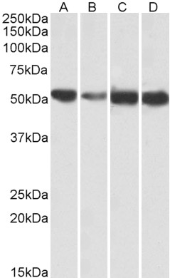
ATP5A1 Antibody (internal region, near N-Term)
Peptide-affinity purified goat antibody
- SPECIFICATION
- CITATIONS
- PROTOCOLS
- BACKGROUND

Application
| WB, E |
|---|---|
| Primary Accession | P25705 |
| Other Accession | NP_004037.1, NP_001244263.1, NP_001001935.1, 498, 11946 (mouse), 65262 (rat) |
| Reactivity | Human, Mouse, Rat, Pig |
| Predicted | Dog, Cow |
| Host | Goat |
| Clonality | Polyclonal |
| Concentration | 0.5 mg/ml |
| Isotype | IgG |
| Calculated MW | 59751 Da |
| Gene ID | 498 |
|---|---|
| Other Names | ATP synthase subunit alpha, mitochondrial, ATP5A1, ATP5A, ATP5AL2, ATPM |
| Format | 0.5 mg/ml in Tris saline, 0.02% sodium azide, pH7.3 with 0.5% bovine serum albumin |
| Storage | Maintain refrigerated at 2-8°C for up to 6 months. For long term storage store at -20°C in small aliquots to prevent freeze-thaw cycles. |
| Precautions | ATP5A1 Antibody (internal region, near N-Term) is for research use only and not for use in diagnostic or therapeutic procedures. |
| Name | ATP5F1A (HGNC:823) |
|---|---|
| Function | Mitochondrial membrane ATP synthase (F(1)F(0) ATP synthase or Complex V) produces ATP from ADP in the presence of a proton gradient across the membrane which is generated by electron transport complexes of the respiratory chain. F-type ATPases consist of two structural domains, F(1) - containing the extramembraneous catalytic core, and F(0) - containing the membrane proton channel, linked together by a central stalk and a peripheral stalk. During catalysis, ATP synthesis in the catalytic domain of F(1) is coupled via a rotary mechanism of the central stalk subunits to proton translocation. Subunits alpha and beta form the catalytic core in F(1). Rotation of the central stalk against the surrounding alpha(3)beta(3) subunits leads to hydrolysis of ATP in three separate catalytic sites on the beta subunits. Subunit alpha does not bear the catalytic high-affinity ATP-binding sites (By similarity). Binds the bacterial siderophore enterobactin and can promote mitochondrial accumulation of enterobactin-derived iron ions (PubMed:30146159). |
| Cellular Location | Mitochondrion. Mitochondrion inner membrane {ECO:0000250|UniProtKB:P19483}; Peripheral membrane protein {ECO:0000250|UniProtKB:P19483}; Matrix side {ECO:0000250|UniProtKB:P19483}. Cell membrane; Peripheral membrane protein; Extracellular side. Note=Colocalizes with HRG on the cell surface of T-cells (PubMed:19285951). |
| Tissue Location | Fetal lung, heart, liver, gut and kidney. Expressed at higher levels in the fetal brain, retina and spinal cord |

Thousands of laboratories across the world have published research that depended on the performance of antibodies from Abcepta to advance their research. Check out links to articles that cite our products in major peer-reviewed journals, organized by research category.
info@abcepta.com, and receive a free "I Love Antibodies" mug.

Provided below are standard protocols that you may find useful for product applications.
Background
This antibody is expected to recognize all reported isoforms (NP_004037.1; NP_001244263.1; NP_001001935.1). Reported variants represent identical protein: NP_001244264.1, NP_001001935.1. Reported variants represent identical protein: NP_004037.1, NP_00100
References
The putative tumour modifier gene ATP5A1 is not mutated in human colorectal cancer cell lines but expression levels correlate with TP53 mutations and chromosomal instability. Seth R, Keeley J, Abu-Ali G, Crook S, Jackson D, Ilyas M. Journal of clinical pathology 2009 Jul 62 (7): 598-603. PMID: 19261598
If you have used an Abcepta product and would like to share how it has performed, please click on the "Submit Review" button and provide the requested information. Our staff will examine and post your review and contact you if needed.
If you have any additional inquiries please email technical services at tech@abcepta.com.
Ordering Information
Other Products
Shipping Information
