


 Foundational characteristics of cancer include proliferation, angiogenesis, migration, evasion of apoptosis, and cellular immortality. Find key markers for these cellular processes and antibodies to detect them.
 The SUMOplot™ Analysis Program predicts and scores sumoylation sites in your protein. SUMOylation is a post-translational modification involved in various cellular processes, such as nuclear-cytosolic transport, transcriptional regulation, apoptosis, protein stability, response to stress, and progression through the cell cycle.
 The Autophagy Receptor Motif Plotter predicts and scores autophagy receptor binding sites in your protein. Identifying proteins connected to this pathway is critical to understanding the role of autophagy in physiological as well as pathological processes such as development, differentiation, neurodegenerative diseases, stress, infection, and cancer.
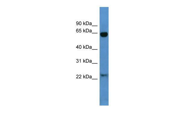
Baiap2 antibody - N-terminal region
Rabbit Polyclonal Antibody
- SPECIFICATION
- CITATIONS
- PROTOCOLS
- BACKGROUND

Application
| WB |
|---|---|
| Primary Accession | Q8BKX1 |
| Other Accession | NM_001037755, NP_001032844 |
| Reactivity | Human, Mouse, Rat, Pig, Horse, Bovine, Dog |
| Predicted | Human, Mouse, Rat, Chicken, Bovine, Guinea Pig |
| Host | Rabbit |
| Clonality | Polyclonal |
| Calculated MW | 59kDa |
| Gene ID | 108100 |
|---|---|
| Alias Symbol | IRSp53, R75030 |
| Other Names | Brain-specific angiogenesis inhibitor 1-associated protein 2, BAI-associated protein 2, BAI1-associated protein 2, Insulin receptor substrate protein of 53 kDa, IRSp53, Insulin receptor substrate p53, Insulin receptor tyrosine kinase 53 kDa substrate, Baiap2 |
| Format | Liquid. Purified antibody supplied in 1x PBS buffer with 0.09% (w/v) sodium azide and 2% sucrose. |
| Reconstitution & Storage | Add 50 ul of distilled water. Final anti-Baiap2 antibody concentration is 1 mg/ml in PBS buffer with 2% sucrose. For longer periods of storage, store at 20°C. Avoid repeat freeze-thaw cycles. |
| Precautions | Baiap2 antibody - N-terminal region is for research use only and not for use in diagnostic or therapeutic procedures. |
| Name | Baiap2 |
|---|---|
| Function | Adapter protein that links membrane-bound small G-proteins to cytoplasmic effector proteins. Necessary for CDC42-mediated reorganization of the actin cytoskeleton and for RAC1-mediated membrane ruffling. Involved in the regulation of the actin cytoskeleton by WASF family members and the Arp2/3 complex. Plays a role in neurite growth. Acts syngeristically with ENAH to promote filipodia formation. Plays a role in the reorganization of the actin cytoskeleton in response to bacterial infection. Participates in actin bundling when associated with EPS8, promoting filopodial protrusions. |
| Cellular Location | Cytoplasm. Membrane; Peripheral membrane protein. Cell projection, filopodium. Cell projection, ruffle. Cytoplasm, cytoskeleton. Note=Detected throughout the cytoplasm in the absence of specific binding partners. Detected in filopodia and close to membrane ruffles. Recruited to actin pedestals that are formed upon infection by bacteria at bacterial attachment sites (By similarity). |
| Tissue Location | Detected in liver, brain, olfactory bulb, brain cortex, caudate putamen, hypothalamus and cerebellum |
Research Areas
Citations (0)

Thousands of laboratories across the world have published research that depended on the performance of antibodies from Abcepta to advance their research. Check out links to articles that cite our products in major peer-reviewed journals, organized by research category.
Submit your citation using an Abcepta antibody to
info@abcepta.com, and receive a free "I Love Antibodies" mug.
info@abcepta.com, and receive a free "I Love Antibodies" mug.

Application Protocols
Provided below are standard protocols that you may find useful for product applications.
Abcepta welcomes feedback from its customers.
If you have used an Abcepta product and would like to share how it has performed, please click on the "Submit Review" button and provide the requested information. Our staff will examine and post your review and contact you if needed.
If you have any additional inquiries please email technical services at tech@abcepta.com.
$ 389.00
Cat# AI10183
Ordering Information
United States
AlbaniaAustraliaAustriaBelgiumBosnia & HerzegovinaBrazilBulgariaCanadaCentral AmericaChinaCroatiaCyprusCzech RepublicDenmarkEstoniaFinlandFranceGermanyGreeceHong KongHungaryIcelandIndiaIndonesiaIrelandIsraelItalyJapanLatviaLithuaniaLuxembourgMacedoniaMalaysiaMaltaNetherlandsNew ZealandNorwayPakistanPolandPortugalRomaniaSerbiaSingaporeSlovakiaSloveniaSouth AfricaSouth KoreaSpainSwedenSwitzerlandTaiwanTurkeyUnited KingdomUnited StatesVietnamWorldwideOthers
USA Headquarters
(888) 735-7227 / (858) 622-0099 or (858) 875-1900
Other Products
Shipping Information
Domestic orders (in stock items)
Shipped out the same day. Orders placed after 1 PM (PST) will ship out the next business day.
International orders
Contact your local distributors
