


 Foundational characteristics of cancer include proliferation, angiogenesis, migration, evasion of apoptosis, and cellular immortality. Find key markers for these cellular processes and antibodies to detect them.
 The SUMOplot™ Analysis Program predicts and scores sumoylation sites in your protein. SUMOylation is a post-translational modification involved in various cellular processes, such as nuclear-cytosolic transport, transcriptional regulation, apoptosis, protein stability, response to stress, and progression through the cell cycle.
 The Autophagy Receptor Motif Plotter predicts and scores autophagy receptor binding sites in your protein. Identifying proteins connected to this pathway is critical to understanding the role of autophagy in physiological as well as pathological processes such as development, differentiation, neurodegenerative diseases, stress, infection, and cancer.
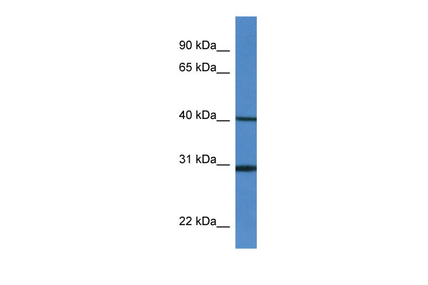
H13 antibody - middle region
Rabbit Polyclonal Antibody
- SPECIFICATION
- CITATIONS
- PROTOCOLS
- BACKGROUND

Application
| WB |
|---|---|
| Primary Accession | Q9D8V0 |
| Other Accession | NM_010376, NP_034506 |
| Reactivity | Human, Mouse, Rat, Rabbit, Pig, Horse, Bovine, Guinea Pig, Dog |
| Predicted | Mouse |
| Host | Rabbit |
| Clonality | Polyclonal |
| Calculated MW | 42kDa |
| Gene ID | 14950 |
|---|---|
| Alias Symbol | 1200006O09Rik, 4930443L17Rik, 5031424B04Rik, AV020344, H-13, Hm13, PSL3, Spp |
| Other Names | Minor histocompatibility antigen H13, 3.4.23.-, Presenilin-like protein 3, Signal peptide peptidase, Hm13, H13, Psl3 |
| Format | Liquid. Purified antibody supplied in 1x PBS buffer with 0.09% (w/v) sodium azide and 2% sucrose. |
| Reconstitution & Storage | Add 50 ul of distilled water. Final anti-H13 antibody concentration is 1 mg/ml in PBS buffer with 2% sucrose. For longer periods of storage, store at 20°C. Avoid repeat freeze-thaw cycles. |
| Precautions | H13 antibody - middle region is for research use only and not for use in diagnostic or therapeutic procedures. |
| Name | Hm13 |
|---|---|
| Synonyms | H13, Psl3 |
| Function | Catalyzes intramembrane proteolysis of some signal peptides after they have been cleaved from a preprotein, resulting in the release of the fragment from the ER membrane into the cytoplasm. Required to generate lymphocyte cell surface (HLA-E) epitopes derived from MHC class I signal peptides. Involved in the intramembrane cleavage of the integral membrane protein PSEN1. Cleaves the integral membrane protein XBP1 isoform 1 in a DERL1/RNF139-dependent manner (By similarity). May play a role in graft rejection (PubMed:9354467). |
| Cellular Location | Endoplasmic reticulum membrane; Multi-pass membrane protein. Membrane {ECO:0000250|UniProtKB:Q8TCT9}; Multi-pass membrane protein {ECO:0000250|UniProtKB:Q8TCT9}; Lumenal side {ECO:0000250|UniProtKB:Q8TCT9} |
| Tissue Location | Widely expressed with highest levels in liver and kidney. In the brain, expressed predominantly in hippocampus, amygdala, piriform cortex, choroid plexus and arcuate nucleus of the hypothalamic area. Isoform 1 is more strongly expressed than isoform 4 in most tissues except brain and skeletal muscle where isoform 4 is the dominant isoform and in testis where isoform 1 and isoform 4 are expressed at similar levels. In the brain, isoform 4 is not detected in the choroid plexus. |

Thousands of laboratories across the world have published research that depended on the performance of antibodies from Abcepta to advance their research. Check out links to articles that cite our products in major peer-reviewed journals, organized by research category.
info@abcepta.com, and receive a free "I Love Antibodies" mug.

Provided below are standard protocols that you may find useful for product applications.
References
Urny J.,et al.Gene Expr. Patterns 3:685-691(2003).
Urny J.,et al.Biochim. Biophys. Acta 1759:159-165(2006).
Irmler M.,et al.Submitted (SEP-2001) to the EMBL/GenBank/DDBJ databases.
Brown A.C.,et al.Submitted (FEB-2002) to the EMBL/GenBank/DDBJ databases.
Carninci P.,et al.Science 309:1559-1563(2005).
If you have used an Abcepta product and would like to share how it has performed, please click on the "Submit Review" button and provide the requested information. Our staff will examine and post your review and contact you if needed.
If you have any additional inquiries please email technical services at tech@abcepta.com.
Ordering Information
Other Products
Shipping Information
