


 Foundational characteristics of cancer include proliferation, angiogenesis, migration, evasion of apoptosis, and cellular immortality. Find key markers for these cellular processes and antibodies to detect them.
 The SUMOplot™ Analysis Program predicts and scores sumoylation sites in your protein. SUMOylation is a post-translational modification involved in various cellular processes, such as nuclear-cytosolic transport, transcriptional regulation, apoptosis, protein stability, response to stress, and progression through the cell cycle.
 The Autophagy Receptor Motif Plotter predicts and scores autophagy receptor binding sites in your protein. Identifying proteins connected to this pathway is critical to understanding the role of autophagy in physiological as well as pathological processes such as development, differentiation, neurodegenerative diseases, stress, infection, and cancer.
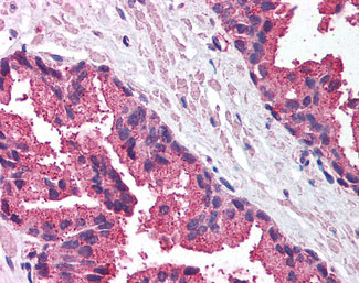
MICA Antibody (C-Terminus)
Rabbit Polyclonal Antibody
- SPECIFICATION
- CITATIONS
- PROTOCOLS
- BACKGROUND

Application
| WB, IHC-P |
|---|---|
| Primary Accession | Q29983 |
| Reactivity | Human, Mouse |
| Host | Rabbit |
| Clonality | Polyclonal |
| Calculated MW | 43kDa |
| Dilution | IHC-P (5 µg/ml), WB (0.5-1 µg/ml), |
| Gene ID | 100507436 |
|---|---|
| Other Names | MHC class I polypeptide-related sequence A, MIC-A, MICA {ECO:0000312|EMBL:CAI41907.1} |
| Target/Specificity | a 16 amino acid peptide from near the carboxy terminus of human MICA |
| Reconstitution & Storage | Short term 4°C, long term aliquot and store at -20°C, avoid freeze thaw cycles. Store undiluted. |
| Precautions | MICA Antibody (C-Terminus) is for research use only and not for use in diagnostic or therapeutic procedures. |
| Name | MICA {ECO:0000312|EMBL:CAI41907.1} |
|---|---|
| Function | Seems to have no role in antigen presentation. Acts as a stress-induced self-antigen that is recognized by gamma delta T-cells. Ligand for the KLRK1/NKG2D receptor. Binding to KLRK1 leads to cell lysis. |
| Cellular Location | Cell membrane; Single-pass type I membrane protein. Cytoplasm. Note=Expressed on the cell surface in gastric epithelium, endothelial cells and fibroblasts and in the cytoplasm in keratinocytes and monocytes. Infection with human adenovirus 5 suppresses cell surface expression due to the adenoviral E3-19K protein which causes retention in the endoplasmic reticulum |
| Tissue Location | Widely expressed with the exception of the central nervous system where it is absent. Expressed predominantly in gastric epithelium and also in monocytes, keratinocytes, endothelial cells, fibroblasts and in the outer layer of Hassal's corpuscles within the medulla of normal thymus. In skin, expressed mainly in the keratin layers, basal cells, ducts and follicles. Also expressed in many, but not all, epithelial tumors of lung, breast, kidney, ovary, prostate and colon. In thyomas, overexpressed in cortical and medullar epithelial cells. Tumors expressing MICA display increased levels of gamma delta T-cells. |

Thousands of laboratories across the world have published research that depended on the performance of antibodies from Abcepta to advance their research. Check out links to articles that cite our products in major peer-reviewed journals, organized by research category.
info@abcepta.com, and receive a free "I Love Antibodies" mug.

Provided below are standard protocols that you may find useful for product applications.
Background
Seems to have no role in antigen presentation. Acts as a stress-induced self-antigen that is recognized by gamma delta T- cells. Ligand for the KLRK1/NKG2D receptor. Binding to KLRK1 leads to cell lysis.
References
Bahram S.,et al.Proc. Natl. Acad. Sci. U.S.A. 91:6259-6263(1994).
Bahram S.,et al.Immunogenetics 44:80-81(1996).
Mungall A.J.,et al.Nature 425:805-811(2003).
Leelayuwat C.,et al.Immunogenetics 40:339-351(1994).
Fodil N.,et al.Immunogenetics 44:351-357(1996).
If you have used an Abcepta product and would like to share how it has performed, please click on the "Submit Review" button and provide the requested information. Our staff will examine and post your review and contact you if needed.
If you have any additional inquiries please email technical services at tech@abcepta.com.
Ordering Information
Other Products
Shipping Information
