


 Foundational characteristics of cancer include proliferation, angiogenesis, migration, evasion of apoptosis, and cellular immortality. Find key markers for these cellular processes and antibodies to detect them.
 The SUMOplot™ Analysis Program predicts and scores sumoylation sites in your protein. SUMOylation is a post-translational modification involved in various cellular processes, such as nuclear-cytosolic transport, transcriptional regulation, apoptosis, protein stability, response to stress, and progression through the cell cycle.
 The Autophagy Receptor Motif Plotter predicts and scores autophagy receptor binding sites in your protein. Identifying proteins connected to this pathway is critical to understanding the role of autophagy in physiological as well as pathological processes such as development, differentiation, neurodegenerative diseases, stress, infection, and cancer.
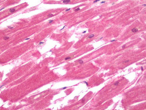
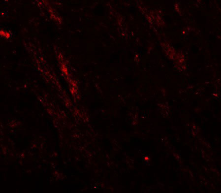
CACNA1H / Cav3.2 Antibody (Internal)
Rabbit Polyclonal Antibody
- SPECIFICATION
- CITATIONS
- PROTOCOLS
- BACKGROUND

Application
| WB, IHC-P, IF, E |
|---|---|
| Primary Accession | O95180 |
| Reactivity | Human, Mouse, Rat |
| Host | Rabbit |
| Clonality | Polyclonal |
| Calculated MW | 259kDa |
| Dilution | IF (20 µg/ml), IHC-P (10 µg/ml), WB (1-2 µg/ml) |
| Gene ID | 8912 |
|---|---|
| Other Names | Voltage-dependent T-type calcium channel subunit alpha-1H, Low-voltage-activated calcium channel alpha1 3.2 subunit, Voltage-gated calcium channel subunit alpha Cav3.2, CACNA1H |
| Target/Specificity | CACNA1H antibody is human, mouse and rat reactive. Multiple isoforms of CACNA1H are known to exist. |
| Reconstitution & Storage | Long term: -20°C; Short term: +4°C. Avoid repeat freeze-thaw cycles. |
| Precautions | CACNA1H / Cav3.2 Antibody (Internal) is for research use only and not for use in diagnostic or therapeutic procedures. |
| Name | CACNA1H (HGNC:1395) |
|---|---|
| Function | Voltage-sensitive calcium channel that gives rise to T-type calcium currents. T-type calcium channels belong to the 'low-voltage activated (LVA)' group. A particularity of this type of channel is an opening at quite negative potentials, and a voltage-dependent inactivation (PubMed:9670923, PubMed:9930755, PubMed:27149520). T-type channels serve pacemaking functions in both central neurons and cardiac nodal cells and support calcium signaling in secretory cells and vascular smooth muscle (Probable). They may also be involved in the modulation of firing patterns of neurons (PubMed:15048902). In the adrenal zona glomerulosa, participates in the signaling pathway leading to aldosterone production in response to either AGT/angiotensin II, or hyperkalemia (PubMed:25907736, PubMed:27729216). |
| Cellular Location | Cell membrane; Multi-pass membrane protein. Note=Interaction with STAC increases expression at the cell membrane. |
| Tissue Location | Expressed in the adrenal glomerulosa (at protein level) (PubMed:25907736, PubMed:27729216). In nonneuronal tissues, the highest expression levels are found in the kidney, liver, and heart. In the brain, most abundant in the amygdala, caudate nucleus, and putamen (PubMed:9670923, PubMed:9930755). In the heart, expressed in blood vessels. [Isoform 2]: Expressed in testis, primarily in the germ cells, but not in other portions of the reproductive tract, such as ductus deferens (PubMed:11751928). Not expressed in the brain (PubMed:11751928). |

Thousands of laboratories across the world have published research that depended on the performance of antibodies from Abcepta to advance their research. Check out links to articles that cite our products in major peer-reviewed journals, organized by research category.
info@abcepta.com, and receive a free "I Love Antibodies" mug.

Provided below are standard protocols that you may find useful for product applications.
Background
Voltage-sensitive calcium channels (VSCC) mediate the entry of calcium ions into excitable cells and are also involved in a variety of calcium-dependent processes, including muscle contraction, hormone or neurotransmitter release, gene expression, cell motility, cell division and cell death. The isoform alpha-1H gives rise to T-type calcium currents. T-type calcium channels belong to the "low-voltage activated (LVA)" group and are strongly blocked by nickel and mibefradil. A particularity of this type of channels is an opening at quite negative potentials, and a voltage-dependent inactivation. T-type channels serve pacemaking functions in both central neurons and cardiac nodal cells and support calcium signaling in secretory cells and vascular smooth muscle. They may also be involved in the modulation of firing patterns of neurons which is important for information processing as well as in cell growth processes.
References
Cribbs L.L.,et al.Circ. Res. 83:103-109(1998).
Cribbs L.L.,et al.Submitted (JUL-2001) to the EMBL/GenBank/DDBJ databases.
Williams M.E.,et al.J. Neurochem. 72:791-799(1999).
Jagannathan S.,et al.J. Biol. Chem. 277:8449-8456(2002).
Daniels R.J.,et al.Hum. Mol. Genet. 10:339-352(2001).
If you have used an Abcepta product and would like to share how it has performed, please click on the "Submit Review" button and provide the requested information. Our staff will examine and post your review and contact you if needed.
If you have any additional inquiries please email technical services at tech@abcepta.com.
Ordering Information
Other Products
Shipping Information
