


 Foundational characteristics of cancer include proliferation, angiogenesis, migration, evasion of apoptosis, and cellular immortality. Find key markers for these cellular processes and antibodies to detect them.
 The SUMOplot™ Analysis Program predicts and scores sumoylation sites in your protein. SUMOylation is a post-translational modification involved in various cellular processes, such as nuclear-cytosolic transport, transcriptional regulation, apoptosis, protein stability, response to stress, and progression through the cell cycle.
 The Autophagy Receptor Motif Plotter predicts and scores autophagy receptor binding sites in your protein. Identifying proteins connected to this pathway is critical to understanding the role of autophagy in physiological as well as pathological processes such as development, differentiation, neurodegenerative diseases, stress, infection, and cancer.
EDA / ED1 Antibody (Internal)
Rabbit Polyclonal Antibody

- SPECIFICATION
- CITATIONS
- PROTOCOLS
- BACKGROUND

Application
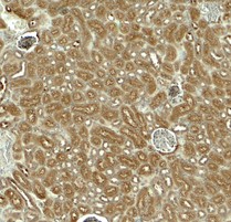
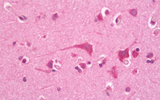
| WB, IHC-P, IF, E |
|---|---|
| Primary Accession | Q92838 |
| Other Accession | 1896 |
| Reactivity | Human, Mouse, Rat |
| Host | Rabbit |
| Clonality | Polyclonal |
| Calculated MW | 41294 Da |
| Dilution | IF (20 µg/ml), IHC-P (10 µg/ml), WB (1 - 2 µg/ml), |
| Gene ID | 1896 |
|---|---|
| Other Names | EDA, ECTD1, Ectodermal dysplasia protein, Ectodysplasin-A, ED1, ED1-A1, Ectodysplasin A, EDA protein, EDA1, EDA2, Hed, HED1, ODT1, STHAGX1, XHED, XLHED, ED1-A2, Oligodontia 1 |
| Target/Specificity | EDA1 antibody is human, mouse and rat reactive. Multiple isoforms of EDA1 are known to exist. |
| Reconstitution & Storage | PBS, 0.02% sodium azide. Long term: -20°C; Short term: +4°C. Avoid repeat freeze-thaw cycles. |
| Precautions | EDA / ED1 Antibody (Internal) is for research use only and not for use in diagnostic or therapeutic procedures. |
| Name | EDA |
|---|---|
| Synonyms | ED1, EDA2 |
| Function | Cytokine which is involved in epithelial-mesenchymal signaling during morphogenesis of ectodermal organs. Functions as a ligand activating the DEATH-domain containing receptors EDAR and EDA2R (PubMed:8696334, PubMed:11039935, PubMed:27144394, PubMed:34582123). May also play a role in cell adhesion (By similarity). |
| Cellular Location | Cell membrane {ECO:0000250|UniProtKB:O54693}; Single-pass type II membrane protein {ECO:0000250|UniProtKB:O54693} |
| Tissue Location | Not abundant; expressed in specific cell types of ectodermal (but not mesodermal) origin of keratinocytes, hair follicles, sweat glands. Also in adult heart, liver, muscle, pancreas, prostate, fetal liver, uterus, small intestine and umbilical chord {ECO:0000269|Ref.6} |

Thousands of laboratories across the world have published research that depended on the performance of antibodies from Abcepta to advance their research. Check out links to articles that cite our products in major peer-reviewed journals, organized by research category.
info@abcepta.com, and receive a free "I Love Antibodies" mug.

Provided below are standard protocols that you may find useful for product applications.
Background
Seems to be involved in epithelial-mesenchymal signaling during morphogenesis of ectodermal organs. Isoform 1 binds only to the receptor EDAR, while isoform 3 binds exclusively to the receptor XEDAR.
References
Kere J.,et al.Nat. Genet. 13:409-416(1996).
Monreal A.W.,et al.Am. J. Hum. Genet. 63:380-389(1998).
Bayes M.,et al.Hum. Mol. Genet. 7:1661-1669(1998).
Ross M.T.,et al.Nature 434:325-337(2005).
Kobielak K.,et al.Eur. J. Hum. Genet. Suppl. 7:104-104(1999).
If you have used an Abcepta product and would like to share how it has performed, please click on the "Submit Review" button and provide the requested information. Our staff will examine and post your review and contact you if needed.
If you have any additional inquiries please email technical services at tech@abcepta.com.
Ordering Information
Other Products
Shipping Information
