


 Foundational characteristics of cancer include proliferation, angiogenesis, migration, evasion of apoptosis, and cellular immortality. Find key markers for these cellular processes and antibodies to detect them.
 The SUMOplot™ Analysis Program predicts and scores sumoylation sites in your protein. SUMOylation is a post-translational modification involved in various cellular processes, such as nuclear-cytosolic transport, transcriptional regulation, apoptosis, protein stability, response to stress, and progression through the cell cycle.
 The Autophagy Receptor Motif Plotter predicts and scores autophagy receptor binding sites in your protein. Identifying proteins connected to this pathway is critical to understanding the role of autophagy in physiological as well as pathological processes such as development, differentiation, neurodegenerative diseases, stress, infection, and cancer.
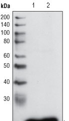
GSTP1 Antibody
Purified Mouse Monoclonal Antibody


- SPECIFICATION
- CITATIONS
- PROTOCOLS
- BACKGROUND

Application
| WB, IHC, FC, ICC, E |
|---|---|
| Primary Accession | P09211 |
| Reactivity | Human |
| Host | Mouse |
| Clonality | Monoclonal |
| Clone Names | 3F2C2 |
| Isotype | IgG1 |
| Calculated MW | 23kDa |
| Description | GSTP1 (glutathione-S-transferase, pi 1), also called GST3/DFN7, is a family of enzymes that play an important role in detoxification by catalyzing the conjugation of many hydrophobic and electrophilic compounds with reduced glutathione. GSTP1 act like a tumor suppressor gene, which when inactivated leads to tumor growth, and the -class glutathione S-transferase is commonly inactivated by somatic CpGisland hypermethylation in cancers of the prostate, liver, and breast. Methylation of regulatory sequences at the GSTP1 gene locus is found in the vast majority (>90%) of prostate carcinomas and is associated with transcriptional down-regulation. |
| Immunogen | Purified recombinant fragment of human GSTP1 expressed in E. Coli. |
| Formulation | Ascitic fluid containing 0.03% sodium azide. |
| Gene ID | 2950 |
|---|---|
| Other Names | Glutathione S-transferase P, 2.5.1.18, GST class-pi, GSTP1-1, GSTP1, FAEES3, GST3 |
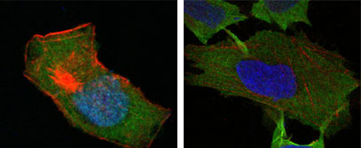
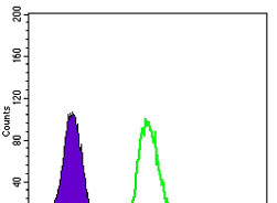
| Dilution | WB~~1/500 - 1/2000 IHC~~1/500 - 1/2000 IF~~1/200 - 1/1000 FC~~1/200 - 1/400 |
| Storage | Maintain refrigerated at 2-8°C for up to 6 months. For long term storage store at -20°C in small aliquots to prevent freeze-thaw cycles. |
| Precautions | GSTP1 Antibody is for research use only and not for use in diagnostic or therapeutic procedures. |
| Name | GSTP1 (HGNC:4638) |
|---|---|
| Synonyms | FAEES3, GST3 |
| Function | Conjugation of reduced glutathione to a wide number of exogenous and endogenous hydrophobic electrophiles. Involved in the formation of glutathione conjugates of both prostaglandin A2 (PGA2) and prostaglandin J2 (PGJ2) (PubMed:9084911). Participates in the formation of novel hepoxilin regioisomers (PubMed:21046276). Negatively regulates CDK5 activity via p25/p35 translocation to prevent neurodegeneration. |
| Cellular Location | Cytoplasm. Mitochondrion. Nucleus. Note=The 83 N-terminal amino acids function as un uncleaved transit peptide, and arginine residues within it are crucial for mitochondrial localization |

Thousands of laboratories across the world have published research that depended on the performance of antibodies from Abcepta to advance their research. Check out links to articles that cite our products in major peer-reviewed journals, organized by research category.
info@abcepta.com, and receive a free "I Love Antibodies" mug.

Provided below are standard protocols that you may find useful for product applications.
References
1. Kimihiko Satoh, Ken Itoh, Masayuki Yamamoto. 2002. Carcinogenesis. 23: 457 - 462.2. Xiaohui Lin,William G. Nelson.2003.Cancer Research. 63: 498-504.
If you have used an Abcepta product and would like to share how it has performed, please click on the "Submit Review" button and provide the requested information. Our staff will examine and post your review and contact you if needed.
If you have any additional inquiries please email technical services at tech@abcepta.com.
Ordering Information
Other Products
Shipping Information
