


 Foundational characteristics of cancer include proliferation, angiogenesis, migration, evasion of apoptosis, and cellular immortality. Find key markers for these cellular processes and antibodies to detect them.
 The SUMOplot™ Analysis Program predicts and scores sumoylation sites in your protein. SUMOylation is a post-translational modification involved in various cellular processes, such as nuclear-cytosolic transport, transcriptional regulation, apoptosis, protein stability, response to stress, and progression through the cell cycle.
 The Autophagy Receptor Motif Plotter predicts and scores autophagy receptor binding sites in your protein. Identifying proteins connected to this pathway is critical to understanding the role of autophagy in physiological as well as pathological processes such as development, differentiation, neurodegenerative diseases, stress, infection, and cancer.
AURKA Antibody
Purified Mouse Monoclonal Antibody
- SPECIFICATION
- CITATIONS
- PROTOCOLS
- BACKGROUND

Application
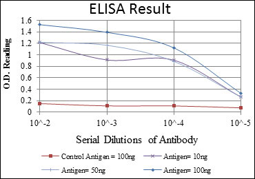
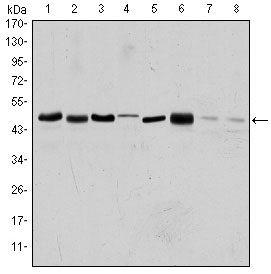
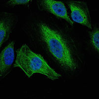
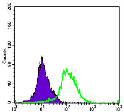
| WB, FC, ICC, E |
|---|---|
| Primary Accession | O14965 |
| Reactivity | Human, Rat, Monkey |
| Host | Mouse |
| Clonality | Monoclonal |
| Clone Names | 1F8 |
| Isotype | IgG1 |
| Calculated MW | 48kDa |
| Description | Aurora A plays a role in cell cycle regulation during anaphase and/or telophase, in relation to the function of the centrosome/spindle pole region during chromosome segregation. Aurora A plays a key role during tumor development and progression and is overexpressed in many human cancers including breast, ovarian and colorectal. Aurora A is viewed as a potential target for anticancer drug treatment.Tissue specificity: Highly expressed in testis and weakly in skeletal muscle, thymus and spleen. Also highly expressed in colon, ovarian, prostate, neuroblastoma, breast and cervical cancer cell lines. |
| Immunogen | Purified recombinant fragment of human AURKA expressed in E. Coli. |
| Formulation | Ascitic fluid containing 0.03% sodium azide. |
| Gene ID | 6790 |
|---|---|
| Other Names | Aurora kinase A, 2.7.11.1, Aurora 2, Aurora/IPL1-related kinase 1, ARK-1, Aurora-related kinase 1, hARK1, Breast tumor-amplified kinase, Serine/threonine-protein kinase 15, Serine/threonine-protein kinase 6, Serine/threonine-protein kinase aurora-A, AURKA |
| Dilution | E~~1/10000 WB~~1/500 - 1/2000 IF~~1/200 - 1/1000 FC~~1/200 - 1/400 |
| Storage | Maintain refrigerated at 2-8°C for up to 6 months. For long term storage store at -20°C in small aliquots to prevent freeze-thaw cycles. |
| Precautions | AURKA Antibody is for research use only and not for use in diagnostic or therapeutic procedures. |
| Name | AURKA (HGNC:11393) |
|---|---|
| Function | Mitotic serine/threonine kinase that contributes to the regulation of cell cycle progression (PubMed:26246606, PubMed:12390251, PubMed:18615013, PubMed:11039908, PubMed:17125279, PubMed:17360485). Associates with the centrosome and the spindle microtubules during mitosis and plays a critical role in various mitotic events including the establishment of mitotic spindle, centrosome duplication, centrosome separation as well as maturation, chromosomal alignment, spindle assembly checkpoint, and cytokinesis (PubMed:26246606, PubMed:14523000). Required for normal spindle positioning during mitosis and for the localization of NUMA1 and DCTN1 to the cell cortex during metaphase (PubMed:27335426). Required for initial activation of CDK1 at centrosomes (PubMed:13678582, PubMed:15128871). Phosphorylates numerous target proteins, including ARHGEF2, BORA, BRCA1, CDC25B, DLGP5, HDAC6, KIF2A, LATS2, NDEL1, PARD3, PPP1R2, PLK1, RASSF1, TACC3, p53/TP53 and TPX2 (PubMed:18056443, PubMed:15128871, PubMed:14702041, PubMed:11551964, PubMed:15147269, PubMed:15987997, PubMed:17604723, PubMed:18615013). Regulates KIF2A tubulin depolymerase activity (PubMed:19351716). Important for microtubule formation and/or stabilization (PubMed:18056443). Required for normal axon formation (PubMed:19812038). Plays a role in microtubule remodeling during neurite extension (PubMed:19668197). Also acts as a key regulatory component of the p53/TP53 pathway, and particularly the checkpoint- response pathways critical for oncogenic transformation of cells, by phosphorylating and destabilizing p53/TP53 (PubMed:14702041). Phosphorylates its own inhibitors, the protein phosphatase type 1 (PP1) isoforms, to inhibit their activity (PubMed:11551964). Inhibits cilia outgrowth (By similarity). Required for cilia disassembly via phosphorylation of HDAC6 and subsequent deacetylation of alpha-tubulin (PubMed:17604723, PubMed:20643351). Regulates protein levels of the anti-apoptosis protein BIRC5 by suppressing the expression of the SCF(FBXL7) E3 ubiquitin-protein ligase substrate adapter FBXL7 through the phosphorylation of the transcription factor FOXP1 (PubMed:28218735). |
| Cellular Location | Cytoplasm, cytoskeleton, microtubule organizing center, centrosome. Cytoplasm, cytoskeleton, spindle pole. Cytoplasm, cytoskeleton, microtubule organizing center, centrosome, centriole {ECO:0000250|UniProtKB:P97477}. Cell projection, neuron projection {ECO:0000250|UniProtKB:P97477}. Cell projection, cilium. Cytoplasm, cytoskeleton, cilium basal body. Basolateral cell membrane {ECO:0000250|UniProtKB:F1PNY0}. Note=Detected at the neurite hillock in developing neurons (By similarity). Localizes at the centrosome in mitotic cells from early prophase until telophase, but also localizes to the spindle pole MTs from prophase to anaphase (PubMed:9606188, PubMed:17229885, PubMed:21225229). Colocalized with SIRT2 at centrosome (PubMed:22014574). Moves to the midbody during both telophase and cytokinesis (PubMed:17726514). Associates with both the pericentriolar material (PCM) and centrioles (PubMed:22014574). The localization to the spindle poles is regulated by AAAS (PubMed:26246606) {ECO:0000250|UniProtKB:P97477, ECO:0000269|PubMed:17229885, ECO:0000269|PubMed:17726514, ECO:0000269|PubMed:21225229, ECO:0000269|PubMed:22014574, ECO:0000269|PubMed:26246606, ECO:0000269|PubMed:9606188} |
| Tissue Location | Highly expressed in testis and weakly in skeletal muscle, thymus and spleen. Also highly expressed in colon, ovarian, prostate, neuroblastoma, breast and cervical cancer cell lines |

Thousands of laboratories across the world have published research that depended on the performance of antibodies from Abcepta to advance their research. Check out links to articles that cite our products in major peer-reviewed journals, organized by research category.
info@abcepta.com, and receive a free "I Love Antibodies" mug.

Provided below are standard protocols that you may find useful for product applications.
References
1. Cell Cycle. 2008 Nov 15;7(22):3525-33. 2. Oncogene. 2008 Nov 20;27(51):6539-49.
If you have used an Abcepta product and would like to share how it has performed, please click on the "Submit Review" button and provide the requested information. Our staff will examine and post your review and contact you if needed.
If you have any additional inquiries please email technical services at tech@abcepta.com.
Ordering Information
Other Products
Shipping Information
