


 Foundational characteristics of cancer include proliferation, angiogenesis, migration, evasion of apoptosis, and cellular immortality. Find key markers for these cellular processes and antibodies to detect them.
 The SUMOplot™ Analysis Program predicts and scores sumoylation sites in your protein. SUMOylation is a post-translational modification involved in various cellular processes, such as nuclear-cytosolic transport, transcriptional regulation, apoptosis, protein stability, response to stress, and progression through the cell cycle.
 The Autophagy Receptor Motif Plotter predicts and scores autophagy receptor binding sites in your protein. Identifying proteins connected to this pathway is critical to understanding the role of autophagy in physiological as well as pathological processes such as development, differentiation, neurodegenerative diseases, stress, infection, and cancer.
TUBB1 Antibody
Purified Mouse Monoclonal Antibody


- SPECIFICATION
- CITATIONS
- PROTOCOLS
- BACKGROUND

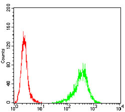
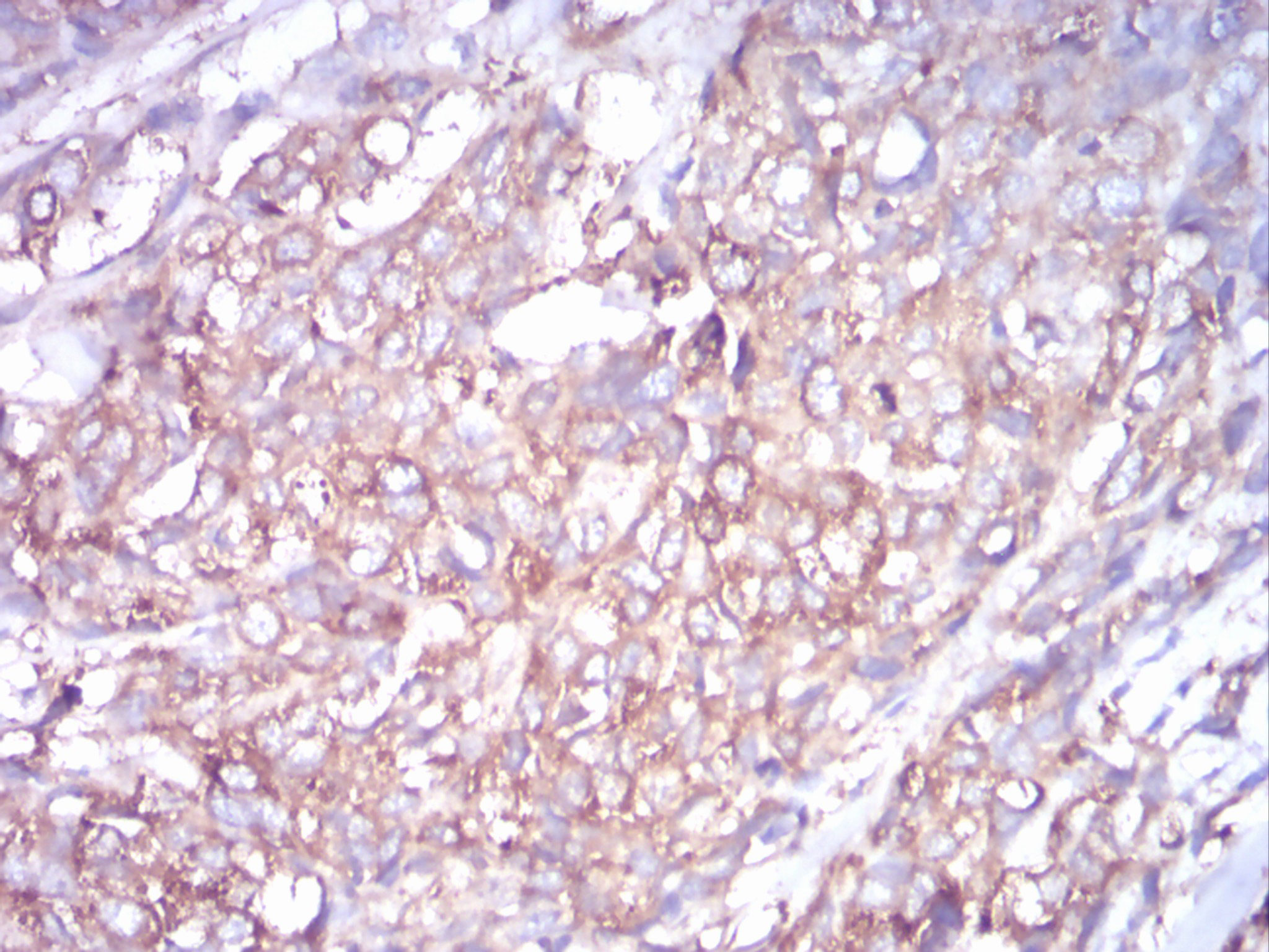
Application
| WB, IHC, FC, ICC, E |
|---|---|
| Primary Accession | Q9H4B7 |
| Reactivity | Human, Mouse, Rat, Monkey |
| Host | Mouse |
| Clonality | Monoclonal |
| Clone Names | 2A1A9 |
| Isotype | IgG1 |
| Calculated MW | 50.3kDa |
| Description | This gene encodes a member of the beta tubulin protein family. Beta tubulins are one of two core protein families (alpha and beta tubulins) that heterodimerize and assemble to form microtubules. This protein is specifically expressed in platelets and megakaryocytes and may be involved in proplatelet production and platelet release. A mutations in this gene is associated with autosomal dominant macrothrombocytopenia. Two pseudogenes of this gene are found on chromosome Y. |
| Immunogen | Purified recombinant fragment of human TUBB1 (AA: 33-166) expressed in E. Coli. |
| Formulation | Purified antibody in PBS with 0.05% sodium azide |
| Gene ID | 81027 |
|---|---|
| Other Names | Tubulin beta-1 chain, TUBB1 |
| Dilution | E~~1/10000 WB~~1/500 - 1/2000 IF~~1/200 - 1/1000 FC~~1/200 - 1/400 IHC~~1/200 - 1/1000 |
| Storage | Maintain refrigerated at 2-8°C for up to 6 months. For long term storage store at -20°C in small aliquots to prevent freeze-thaw cycles. |
| Precautions | TUBB1 Antibody is for research use only and not for use in diagnostic or therapeutic procedures. |
| Name | TUBB1 |
|---|---|
| Function | Tubulin is the major constituent of microtubules, a cylinder consisting of laterally associated linear protofilaments composed of alpha- and beta-tubulin heterodimers. Microtubules grow by the addition of GTP-tubulin dimers to the microtubule end, where a stabilizing cap forms. Below the cap, tubulin dimers are in GDP-bound state, owing to GTPase activity of alpha-tubulin. |
| Cellular Location | Cytoplasm, cytoskeleton |
| Tissue Location | Hematopoietic cell-specific. Major isotype in leukocytes, where it represents 50% of all beta-tubulins |

Thousands of laboratories across the world have published research that depended on the performance of antibodies from Abcepta to advance their research. Check out links to articles that cite our products in major peer-reviewed journals, organized by research category.
info@abcepta.com, and receive a free "I Love Antibodies" mug.

Provided below are standard protocols that you may find useful for product applications.
References
Cancer Res. 2012 Sep 15;72(18):4744-52.Cytoskeleton (Hoboken). 2011 Mar;68(3):175-87.
If you have used an Abcepta product and would like to share how it has performed, please click on the "Submit Review" button and provide the requested information. Our staff will examine and post your review and contact you if needed.
If you have any additional inquiries please email technical services at tech@abcepta.com.
Ordering Information
Other Products
Shipping Information
