


 Foundational characteristics of cancer include proliferation, angiogenesis, migration, evasion of apoptosis, and cellular immortality. Find key markers for these cellular processes and antibodies to detect them.
 The SUMOplot™ Analysis Program predicts and scores sumoylation sites in your protein. SUMOylation is a post-translational modification involved in various cellular processes, such as nuclear-cytosolic transport, transcriptional regulation, apoptosis, protein stability, response to stress, and progression through the cell cycle.
 The Autophagy Receptor Motif Plotter predicts and scores autophagy receptor binding sites in your protein. Identifying proteins connected to this pathway is critical to understanding the role of autophagy in physiological as well as pathological processes such as development, differentiation, neurodegenerative diseases, stress, infection, and cancer.
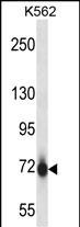
Mouse Pak7 Antibody (N-term)
Affinity Purified Rabbit Polyclonal Antibody (Pab)


- SPECIFICATION
- CITATIONS
- PROTOCOLS
- BACKGROUND

Application
| IHC-P, WB, E |
|---|---|
| Primary Accession | Q8C015 |
| Other Accession | D4A280, NP_766446.2 |
| Reactivity | Human, Mouse |
| Predicted | Rat |
| Host | Rabbit |
| Clonality | Polyclonal |
| Isotype | Rabbit IgG |
| Calculated MW | 80948 Da |
| Antigen Region | 128-157 aa |
| Gene ID | 241656 |
|---|---|
| Other Names | Serine/threonine-protein kinase PAK 7, p21-activated kinase 5, PAK-5, p21-activated kinase 7, PAK-7, Pak7, Pak5 |
| Target/Specificity | This Mouse Pak7 antibody is generated from rabbits immunized with a KLH conjugated synthetic peptide between 128-157 amino acids from the N-terminal region of mouse Pak7. |
| Dilution | WB~~1:1000 IHC-P~~1:10~50 |
| Format | Purified polyclonal antibody supplied in PBS with 0.09% (W/V) sodium azide. This antibody is purified through a protein A column, followed by peptide affinity purification. |
| Storage | Maintain refrigerated at 2-8°C for up to 2 weeks. For long term storage store at -20°C in small aliquots to prevent freeze-thaw cycles. |
| Precautions | Mouse Pak7 Antibody (N-term) is for research use only and not for use in diagnostic or therapeutic procedures. |
| Name | Pak5 {ECO:0000303|PubMed:11756552} |
|---|---|
| Function | Serine/threonine protein kinase that plays a role in a variety of different signaling pathways including cytoskeleton regulation, cell migration, proliferation or cell survival. Activation by various effectors including growth factor receptors or active CDC42 and RAC1 results in a conformational change and a subsequent autophosphorylation on several serine and/or threonine residues. Phosphorylates the proto-oncogene RAF1 and stimulates its kinase activity. Promotes cell survival by phosphorylating the BCL2 antagonist of cell death BAD. Phosphorylates CTNND1, probably to regulate cytoskeletal organization and cell morphology. Keeps microtubules stable through MARK2 inhibition and destabilizes the F-actin network leading to the disappearance of stress fibers and focal adhesions (By similarity). |
| Cellular Location | Mitochondrion. Cytoplasm. Nucleus. Note=Shuttles between the nucleus and the mitochondria, and mitochondrial localization is essential for the role in cell survival. |
| Tissue Location | Highly expressed in brain and eye. Also expressed in adrenal gland, pancreas, prostate and testes. Within the brain, expression is restricted to neurons. Present in brain but not in kidney, lung and spleen (at protein level) |

Thousands of laboratories across the world have published research that depended on the performance of antibodies from Abcepta to advance their research. Check out links to articles that cite our products in major peer-reviewed journals, organized by research category.
info@abcepta.com, and receive a free "I Love Antibodies" mug.

Provided below are standard protocols that you may find useful for product applications.
Background
The activated kinase acts on a variety of targets (By similarity).
References
Gobert, R.P., et al. Mol. Cell. Biol. 29(6):1538-1553(2009)
Nekrasova, T., et al. Dev. Biol. 322(1):95-108(2008)
Pagliarini, D.J., et al. Cell 134(1):112-123(2008)
Sapir, T., et al. J. Neurosci. 28(22):5710-5720(2008)
Trinidad, J.C., et al. Mol. Cell Proteomics 5(5):914-922(2006)
If you have used an Abcepta product and would like to share how it has performed, please click on the "Submit Review" button and provide the requested information. Our staff will examine and post your review and contact you if needed.
If you have any additional inquiries please email technical services at tech@abcepta.com.
Ordering Information
Other Products
Shipping Information
