


 Foundational characteristics of cancer include proliferation, angiogenesis, migration, evasion of apoptosis, and cellular immortality. Find key markers for these cellular processes and antibodies to detect them.
 The SUMOplot™ Analysis Program predicts and scores sumoylation sites in your protein. SUMOylation is a post-translational modification involved in various cellular processes, such as nuclear-cytosolic transport, transcriptional regulation, apoptosis, protein stability, response to stress, and progression through the cell cycle.
 The Autophagy Receptor Motif Plotter predicts and scores autophagy receptor binding sites in your protein. Identifying proteins connected to this pathway is critical to understanding the role of autophagy in physiological as well as pathological processes such as development, differentiation, neurodegenerative diseases, stress, infection, and cancer.
DTNBP1 Antibody (N-term)
Affinity Purified Rabbit Polyclonal Antibody (Pab)

- SPECIFICATION
- CITATIONS
- PROTOCOLS
- BACKGROUND

Application
| WB, E |
|---|---|
| Primary Accession | Q96EV8 |
| Other Accession | NP_898861.1, NP_115498.2 |
| Reactivity | Human |
| Host | Rabbit |
| Clonality | Polyclonal |
| Isotype | Rabbit IgG |
| Calculated MW | 39493 Da |
| Antigen Region | 10-38 aa |
| Gene ID | 84062 |
|---|---|
| Other Names | Dysbindin, Biogenesis of lysosome-related organelles complex 1 subunit 8, BLOC-1 subunit 8, Dysbindin-1, Dystrobrevin-binding protein 1, Hermansky-Pudlak syndrome 7 protein, HPS7 protein, DTNBP1, BLOC1S8 |
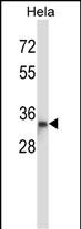
| Target/Specificity | This DTNBP1 antibody is generated from rabbits immunized with a KLH conjugated synthetic peptide between 10-38 amino acids from the N-terminal region of human DTNBP1. |
| Dilution | WB~~1:1000 |
| Format | Purified polyclonal antibody supplied in PBS with 0.09% (W/V) sodium azide. This antibody is purified through a protein A column, followed by peptide affinity purification. |
| Storage | Maintain refrigerated at 2-8°C for up to 2 weeks. For long term storage store at -20°C in small aliquots to prevent freeze-thaw cycles. |
| Precautions | DTNBP1 Antibody (N-term) is for research use only and not for use in diagnostic or therapeutic procedures. |
| Name | DTNBP1 |
|---|---|
| Synonyms | BLOC1S8 |
| Function | Component of the BLOC-1 complex, a complex that is required for normal biogenesis of lysosome-related organelles (LRO), such as platelet dense granules and melanosomes. In concert with the AP-3 complex, the BLOC-1 complex is required to target membrane protein cargos into vesicles assembled at cell bodies for delivery into neurites and nerve terminals. The BLOC-1 complex, in association with SNARE proteins, is also proposed to be involved in neurite extension. Associates with the BLOC-2 complex to facilitate the transport of TYRP1 independent of AP-3 function. Plays a role in synaptic vesicle trafficking and in neurotransmitter release. Plays a role in the regulation of cell surface exposure of DRD2. May play a role in actin cytoskeleton reorganization and neurite outgrowth. May modulate MAPK8 phosphorylation. Appears to promote neuronal transmission and viability through regulating the expression of SNAP25 and SYN1, modulating PI3- kinase-Akt signaling and influencing glutamatergic release. Regulates the expression of SYN1 through binding to its promoter. Modulates prefrontal cortical activity via the dopamine/D2 pathway. |
| Cellular Location | [Isoform 1]: Cytoplasm. Cytoplasmic vesicle membrane; Peripheral membrane protein; Cytoplasmic side. Endosome membrane; Peripheral membrane protein; Cytoplasmic side. Melanosome membrane; Peripheral membrane protein; Cytoplasmic side. Postsynaptic density. Endoplasmic reticulum Nucleus. Note=Mainly cytoplasmic but shuttles between the cytoplasm and nucleus. Exported out of the nucleus via its NES in a XPO1-dependent manner. Nuclear localization is required for regulation of the expression of genes such as SYN1 Detected in neuron cell bodies, axons and dendrites. Mainly located to the postsynaptic density. Detected at tubulovesicular elements in the vicinity of the Golgi apparatus and of melanosomes. Occasionally detected at the membrane of pigmented melanosomes in cultured melanoma cells. The BLOC-1 complex associates with the BLOC-2 complex in early endosome-associated tubules [Isoform 3]: Cytoplasm. Cytoplasmic vesicle membrane; Peripheral membrane protein; Cytoplasmic side. Cytoplasmic vesicle, secretory vesicle, synaptic vesicle membrane; Peripheral membrane protein; Cytoplasmic side. Endosome membrane; Peripheral membrane protein; Cytoplasmic side. Melanosome membrane; Peripheral membrane protein; Cytoplasmic side. Postsynaptic cell membrane. Endoplasmic reticulum Note=Exclusivley cytoplasmic. Predominantly found in the postsynaptic density (PSD). Little association with synaptic vesicles. The BLOC-1 complex associates with the BLOC-2 complex in early endosome-associated tubules. Associated with the AP-3 complex at presynaptic terminals |
| Tissue Location | Detected in brain, in neurons and in neuropil. Isoform 1 is expressed in the cerebral cortex, and hippocampal frontal (HF). Specific expression in the posterior half of the superior temporal gyrus (pSTG). Higher expression of isoform 2 and 3 in the HF than in the pSTG while isoform 1 shows no difference in expression in these areas. In the HF, detected in dentate gyrus (DG) and in pyramidal cells of hippocampus CA2 and CA3 (at protein level). Expressed in all principal neuronal populations of the HF, namely pyramidal neurons in the subiculum and CA1-3, granule cells in the dense cell layer of the DG (DGg), and polymorph cells in the hilus of the DG (DGh). Maximal levels in CA2, CA3, and DGh. Isoform 2 not expressed in the cerebral cortex. |

Thousands of laboratories across the world have published research that depended on the performance of antibodies from Abcepta to advance their research. Check out links to articles that cite our products in major peer-reviewed journals, organized by research category.
info@abcepta.com, and receive a free "I Love Antibodies" mug.

Provided below are standard protocols that you may find useful for product applications.
Background
This gene encodes a protein that may play a role in organelle biogenesis associated with melanosomes, platelet dense granules, and lysosomes. A similar protein in mouse is a component of a protein complex termed biogenesis of lysosome-related organelles complex 1 (BLOC-1), and binds to alpha- and beta-dystrobrevins, which are components of the dystrophin-associated protein complex (DPC). Mutations in this gene are associated with Hermansky-Pudlak syndrome type 7. This gene may also be associated with schizophrenia. Multiple transcript variants encoding distinct isoforms have been identified for this gene.
References
Haukvik, U.K., et al. Prog. Neuropsychopharmacol. Biol. Psychiatry 34(7):1259-1265(2010)
Voisey, J., et al. Eur. Psychiatry 25(6):314-319(2010)
Ruano, G., et al. Pharmacogenomics 11(7):959-971(2010)
Hashimoto, R., et al. Nihon Shinkei Seishin Yakurigaku Zasshi 30(3):103-107(2010)
Voisey, J., et al. Behav Brain Funct 6, 41 (2010) :
If you have used an Abcepta product and would like to share how it has performed, please click on the "Submit Review" button and provide the requested information. Our staff will examine and post your review and contact you if needed.
If you have any additional inquiries please email technical services at tech@abcepta.com.

Ordering Information
Other Products
Shipping Information
