


 Foundational characteristics of cancer include proliferation, angiogenesis, migration, evasion of apoptosis, and cellular immortality. Find key markers for these cellular processes and antibodies to detect them.
 The SUMOplot™ Analysis Program predicts and scores sumoylation sites in your protein. SUMOylation is a post-translational modification involved in various cellular processes, such as nuclear-cytosolic transport, transcriptional regulation, apoptosis, protein stability, response to stress, and progression through the cell cycle.
 The Autophagy Receptor Motif Plotter predicts and scores autophagy receptor binding sites in your protein. Identifying proteins connected to this pathway is critical to understanding the role of autophagy in physiological as well as pathological processes such as development, differentiation, neurodegenerative diseases, stress, infection, and cancer.
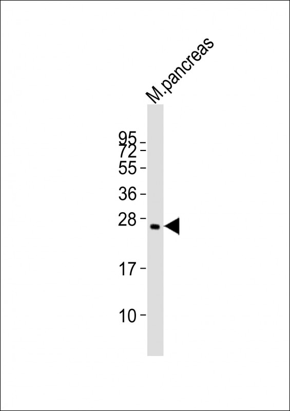
(Mouse) Plet1 Antibody (Center)
Purified Rabbit Polyclonal Antibody (Pab)
- SPECIFICATION
- CITATIONS
- PROTOCOLS
- BACKGROUND

Application
| WB, E |
|---|---|
| Primary Accession | Q8VEN2 |
| Reactivity | Mouse |
| Host | Rabbit |
| Clonality | polyclonal |
| Isotype | Rabbit IgG |
| Calculated MW | 25006 Da |
| Gene ID | 76509 |
|---|---|
| Other Names | Placenta-expressed transcript 1 protein, Antigen mAgK114, Plet1 |
| Target/Specificity | This Mouse Plet1 antibody is generated from a rabbit immunized with a KLH conjugated synthetic peptide between 82-115 amino acids from the Central region of Mouse Plet1. |
| Dilution | WB~~1:2000 |
| Format | Purified polyclonal antibody supplied in PBS with 0.09% (W/V) sodium azide. This antibody is purified through a protein A column, followed by peptide affinity purification. |
| Storage | Maintain refrigerated at 2-8°C for up to 2 weeks. For long term storage store at -20°C in small aliquots to prevent freeze-thaw cycles. |
| Precautions | (Mouse) Plet1 Antibody (Center) is for research use only and not for use in diagnostic or therapeutic procedures. |
| Name | Plet1 |
|---|---|
| Function | Modulates leading keratinocyte migration and cellular adhesion to matrix proteins during a wound-healing response and promotes wound repair. May play a role during trichilemmal differentiation of the hair follicle. |
| Cellular Location | Apical cell membrane; Lipid-anchor, GPI-anchor. Note=Localized at the apical membrane of the most differentiated keratinocytes of the outer root sheath (ORS), clustered mainly in planar regions of the plasma membrane at the base of microvilli |
| Tissue Location | Present in hair follicle cells and sebaceous gland of skin, ciliated epithelial cells of trachea and bronchial tube, striated portion of submandibular gland, distal convoluted tubule cells of kidney, ciliated epithelial cells of oviduct, medulla of adrenal gland and anterior lobe of pituitary gland. Expressed in keratinocytes of the hair follicle at the trichilemmal zone corresponding to the terminally differentiated outermost suprabasal outer root sheath (ORS), including that of the sebaceous gland duct (SGD) and the directly adjacent upper distal end of the companion layer (CL). Expression is similar in all hair follicle growth stages. Also detected during both the early and late anagen phases above the bulge of stem cells Expressed at the leading edge of the epidermal wound. Not expressed in the interfollicular epidermis (IFE), inner root sheath (IRS) and hair fiber. Highly expressed in placenta. Detected in mammary and prostate epithelia and in the pancreas (at protein level) |

Thousands of laboratories across the world have published research that depended on the performance of antibodies from Abcepta to advance their research. Check out links to articles that cite our products in major peer-reviewed journals, organized by research category.
info@abcepta.com, and receive a free "I Love Antibodies" mug.

Provided below are standard protocols that you may find useful for product applications.
Background
Modulates leading keratinocyte migration and cellular adhesion to matrix proteins during a wound-healing response and promotes wound repair. May play a role during trichilemmal differentiation of the hair follicle.
References
Zhao S.-H.,et al.Genomics 84:114-125(2004).
Carninci P.,et al.Science 309:1559-1563(2005).
Takeuchi M.,et al.Zool. Sci. 22:995-1001(2005).
Tatefuji T.,et al.Biol. Pharm. Bull. 29:896-902(2006).
Frankenberg S.,et al.BMC Dev. Biol. 7:8-8(2007).
If you have used an Abcepta product and would like to share how it has performed, please click on the "Submit Review" button and provide the requested information. Our staff will examine and post your review and contact you if needed.
If you have any additional inquiries please email technical services at tech@abcepta.com.
Ordering Information
Other Products
Shipping Information
